BERLIN - Modern kids seem to fall into two categories: those who treat the PlayStation as their BFF (aka best friend forever) and those who live to actually get out and play sports in the real world.
About 35 million children and adolescents in the U.S. participate in organized or recreational sports, said Dr. Kathleen Emery in a talk Saturday at the 2007 International Society for Magnetic Resonance in Medicine (ISMRM) meeting. In general, they concentrate on one sport and train at accelerated levels, she added, resulting in overuse and repetitive stress injuries that are compounded by the very delicate nature of a growing musculoskeletal system.
"I think (pediatric sports injuries) has become one of the most explosive areas that I've seen in terms of MR imaging and it's getting more challenging," Emery said. "Sports are extremely popular and we're seeing more intense training where they don't get breaks (to rest)."
Emery, who is an associate professor of radiology and pediatrics at the Cincinnati Children's Hospital Medical Center in Ohio, discussed patterns of pediatric sports injuries. In a separate presentation, Dr. Diego Jaramillo from Philadelphia offered some typical MR signs to look for in various chronic musculoskeletal issues in the pediatric population.
The weakest link
"There are a lot of things that set the pediatric musculoskeletal system up for injuries that you may not see in adults," Emery said.
The weakest link in the ever-changing pediatric musculoskeletal system is the physis, which is weaker than bone. Subsequently, bone is weaker than ligaments. This weakness occurs in response to puberty and hormonal changes, particularly in boys, Emery said. During the rapid growth of puberty, there is increased stress on tendinous insertions.
While mechanical stress is necessary for normal connective tissue homeostatis, the repetitive loading on young joints, bones, and muscles in sports can actually be harmful. "Sports gives you repetitive, submaximal stress with an occasional and unpredictable maximal load superimposed on top of it," Emery added.
Two of the most common patterns of acute pediatric sports injuries are soft-tissue overuse and growth plate injuries. The former includes Osgood-Schlatter disease and rotator cuff tendinitis. The latter generally takes the form of acute growth plate fractures; about 10% of these physeal injuries are sports-related. It's more common in the upper extremities, she said.
Osseous stress injuries are less common in kids than in adults, but when they turn up, it's in the lower extremities. "The vulnerable period (for osseous stress injuries) is a patient who has take on a new activity, usually within that three-week period after activity initiation where the osteoclastic response in bone exceeds the ability of osteoblastic repair," Emery explained. "The bone scan and MRI are both sensitive but the MR is much more specific."
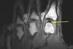
Chronic injuries, which most often heal with activity cessation, include "little league shoulder" in throwing athletes (chronic rotational stress on proximal humeral growth plate) and "gymnast's wrist" from repeated, excess dorsiflexion and axial loading. Emery noted that while MRI may not always be required to make the diagnosis, her sports medicine colleagues do like to see the modality employed as the final word.
"The fact of the matter is that people -- the patient, the parents, the coaches -- want to know. MRI gives them a rapid, graphic confirmation of the diagnosis that is suspected or one that was not suspected," she said. "It allows them to stage the severity of the injury and to determine how long to keep a patient out of sports."
Transformation on MRI
In a separate lecture, Jarmillo outlined what can be seen on various MR sequences when looking at the pediatric musculoskeletal system. Jarmillo is from the University of Pennsylvania and the Children's Hospital of Philadelphia.
As Emery pointed out, the growth plate should be a major focus of MR musculoskeletal exams in a younger population. "The zone of provisional calcification is the area of the newest formation of bone where the cartilage is beginning to mineralize," Jaramillo said. "It's an important landmark because it tells us that the growth plate is healthy."
In terms of specific pulse sequences, cartilage will remain indeterminate on T1-weighted images. Signal from the bone depends on the marrow content, he said. On water-sensitive sequences, the physeal cartilage will be bright, the epiphyseal cartilage will be dark, and the zonal architecture of the cartilage can be seen. On T2-weighted imaging, the physeal cartilage will also be bright. Finally, on gradient-recalled echo (GRE) sequences, bone will be dark and cartilage will be bright, but the zonal architecture of cartilage cannot be seen.
Jaramillo also stressed the importance of the perichondrium, which has a low signal intensity on MRI. "If you have an occult fracture, it's going to show up as a disruption of the perichondrium. It's an important structure to look at," he explained.
Another structure to keep an eye out for is epiphyseal vascular canals. These channels come about when "the cartilage and the (blood) vessels don't speak to each other so the only way that the cartilage can be vascularized is by these canals that go through the cartilage," he said. "You can see those canals because they fill up with gadolinium for many minutes. They are a good indicator of epiphyseal perfusion. They are more prominent with arthritis."
In terms of marrow, Jaramillo said that imaging experts must be sure to separate hematopoietic marrow from disease. This red marrow will have a higher signal than muscle on T1- and T2-weighted imaging and a signal that is equal to muscle on STIR sequences.
"We see this transformation on MRI from red to yellow (fatty) marrow. How do you see hematopoietic marrow form disease? Typically, hematopoietic marrow has these very straight lines (on MR). Disease wouldn't do that."
Finally, Jaramillo discussed the most common indications for gadolinium-enhanced MRI in a pediatric population. In cases of suspected osteomyelitis, if gadolinium uptake does not occur, chances are that antibiotics uptake will not take place either, he said. Contrast-enhanced MR can also be helpful for quantifying necrosis and vascular malformation in tumors. The modality can be used for arthritis, epiphyseal ischemia, and indirect MR arthrography.
Jaramillo highlighted one area in which MRI can be particularly useful: extension of a fracture into unossified epiphysis. In some cases, the fracture will go from the lateral metaphysis of the humerus head into the cartilage. In others, it will stop short of the cartilage, he explained, adding that a fracture that's made its way into the cartilage will require surgical management. However, if the fracture ends before the cartilage, then conservative treatment should suffice, he noted.
In terms of the future of pediatric MR in the musculoskeletal system, Jaramillo said that T2 mapping of the biochemical analysis of cartilage was on the horizon. Emery said that her sports medicine colleagues were looking forward to functional joint imaging.
By Shalmali Pal
AuntMinnie.com staff writer
May 20, 2007
Related Reading
MR spots reason for chronic groin pain, but may not alter management, April 27, 2007
Anatomic variants of the ankle made clear on MRI, March 16, 2007
Some female athletes prone to develop leg pain, September 28, 2006
MRI, US take on diagnostic challenge of PAI in soccer players, August 8, 2006
US overcomes x-ray's limits in pediatric ankle fractures, January 23, 2006
Copyright © 2007 AuntMinnie.com


.fFmgij6Hin.png?auto=compress%2Cformat&fit=crop&h=100&q=70&w=100)

.fFmgij6Hin.png?auto=compress%2Cformat&fit=crop&h=167&q=70&w=250)


