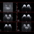
Researchers at the University of Pennsylvania have found a way to coat an iron-based contrast agent for MRI studies so that it only interacts with the acidic environment of tumors, potentially making it safer, cheaper, and more effective than existing alternatives, according to a study published in the journal ACS Nano.
The research was conducted by Andrew Tsourkas, PhD, and graduate student Samuel Crayton of the department of bioengineering in the School of Engineering and Applied Science. It was supported by the U.S. National Institutes of Health and the U.S. Department of Defense Breast Cancer Research Program.
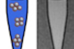
Iron oxide improves MRI images because of its ability to distort the magnetic field of the scanner. Tsourkas and Crayton used glycol chitosan -- a sugar-based polymer that reacts to acids -- to allow the contrast agent nanocarriers to remain neutral when near healthy tissue but to become ionized in low pH. The more malignant a tumor, the more it disrupts surrounding blood vessels, making its environment more acidic; therefore, the glycol chitosan-coated nanoparticles could be good detectors of malignancy, according to Tsourkas and Crayton.


.fFmgij6Hin.png?auto=compress%2Cformat&fit=crop&h=100&q=70&w=100)
.fFmgij6Hin.png?auto=compress%2Cformat&fit=crop&h=167&q=70&w=250)


