An abundance of French charm and research excellence promise to light up McCormick Place in Chicago on Tuesday, when a special plenary session will focus on new frontiers in personalized and integrated oncological approaches.
“French teams have been leading some important studies on personalized care in oncology,” Alain Luciani, MD, PhD, professor of radiology at University Paris Est Creteil (UPEC), told AuntMinnie ahead of the session. “From screening to therapy and now also the follow-up of cancer therapy-induced diseases, we will highlight how both diagnostic and interventional radiology can contribute to optimized patient care and improved outcome.”
 A second-year radiology resident inserts an irreversible electroporation (IRE) needle using Epione, the robotic-assisted platform from Quantum Surgical that supports interventional radiologists in treating inoperable or challenging early-stage tumors. Courtesy of Laurent Milot, MD, PhD.
Advances in interventional radiology, including robot-assisted procedures for abdominal cancers, will be addressed by Laurent Milot, MD, PhD, professor of radiology at the University of Lyon and an interventional radiologist at Edouard Herriot Hospital, Lyon.
“Robotic-assisted interventional radiology procedures are on the rise, theoretically allowing us to better democratize and standardize even the most complex techniques,” he said. “Robotic solutions can also harbor advanced AI-driven planning solutions, potentially improving patient outcomes.”
 Corresponding treatment of a central hepatocellular carcinoma (HCC) shows the precision achieved with the help of the robot, even for junior practitioners. Courtesy of Laurent Milot, MD, PhD.
French radiologists, researchers, and companies are amongst the pioneers in the development and evaluation of robotics in this field, with early research showing very encouraging results, Milot emphasized. Ablation techniques continue to be the mainstay in the treatment of abdominal cancers, he added.
Personalized screening
Tuesday’s RSNA session will also focus on how personalized screening strategies can improve early detection and management of cancer in women, particularly for breast cancers, according to Prof. Isabelle Thomassin-Naggara, head of the Specialized Radiological and Interventional Imaging Service (IRIS) at AP-HP Sorbonne University, Tenon Hospital, Paris.
“France’s breast cancer screening program is evolving to offer personalized follow-up, better coordination, and increased involvement of general practitioners,” she noted. “Innovations include the use of breast tomosynthesis (3D mammography) and digital mammograms for second readings.”
A new risk assessment score has been developed to identify women at higher or lower risk within the average-risk population, and it is being evaluated in the MyPeBS (My Personal Breast Cancer Screening) study. For lower-risk women, the frequency of mammograms may be reduced, while higher-risk women receive intensified surveillance. This approach aims to tailor screening intervals and methods to individual risk profiles, moving beyond age-based screening, she pointed out.
“The role of AI is growing with both detection model or dedication risk-based assessment models, and this will probably improve the definition of intermediate risk women,” Thomassin-Naggara said, adding that an independent platform to evaluate AI algorithms is needed to ensure the implementation of quality of national recommendations. “Women are provided with comprehensive information and decision-support tools, including a dedicated medical consultation at age 50 to discuss screening options and prevention. Clinicians receive training to help women understand the benefits, risks, and limitations of screening, fostering shared decision-making.”
To watch a video interview on the shift to more personalized, risk-adapted, and accessible cancer screening for women, click here.
Radiomics, post-treatment surveillance
The session will also explore how radiomics can leverage developments in quantitative data extracted from images and machine learning.
 Laure Fournier, MD, PhD. Courtesy of Woytek Konarzewski
“Radiomics has moved research from being hypothesis-based to data-driven, and is a way to improve efficiency for imaging biomarker discovery,” said Laure Fournier, MD, PhD, from the Department of Radiology at Georges Pompidou European Hospital in Paris. “The development of radiomics required working on the methodology to improve robustness of results and quality of publications. It is now becoming a mature field. Moreover, working on radiomics allows better understanding of what lies behind deep learning-based methods and applications.”
Her clinical work focuses on urogenital cancers, as well as tumor response to targeted and immunotherapies. Her research in the in vivo imaging lab is centered on radiomics and big data.
Post-treatment surveillance, particularly myocarditis in patients receiving immunotherapy, will be highlighted by Alexis Jacquier, MD, head of radiology at Timone 2 University Hospital in Marseille.
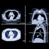
“Immune checkpoint inhibitors (ICIs) have transformed the prognosis of many cancers but can also induce potentially severe adverse effects such as myocarditis,” he told AuntMinnie. Cardiac MRI plays a central role in the diagnosis of this condition. To date, the diagnostic criteria for ICI-associated myocarditis rely largely on those established for viral myocarditis; however, these two diseases differ in several important ways.”
The use of T1 and T2 mapping is particularly important, especially for early detection and for monitoring the progression and healing of ICI-related myocardial inflammation. Optimal management requires a multidisciplinary approach, integrating oncology, cardiology, radiology, and immunology teams to ensure timely diagnosis and safer immunotherapy pathways.
 Patient with diffuse myocardial edema. Myocardial late gadolinium enhancement shows myocarditis caused by immune checkpoint inhibitors. Courtesy of Alexis Jacquier, MD.
During the RSNA session, Marie-France Bellin, MD, president of the French Society of Radiology (SFR), will give an overview of the systemic challenges facing French healthcare and the role radiology plays in addressing them, particularly in cancer care. Luciani will conclude the session by framing the SFR’s strategic vision for radiology, emphasizing international collaboration, innovation, and quality standards. Given the recent strike action and widespread unrest over the 2026 government cuts in medical imaging budgets, this assessment should prove enlightening.
The Country Presents sessions at RSNA 2025 are: T7-RCP03: France Presents: New Frontiers in Personalised and Integrated Oncological Approaches: The French Experience, Tuesday, December 2, 3 p.m. local time; T3-RCP02: United Arab Emirates (UAE) Presents: Radiology at the Forefront: Reflecting the Nation's Progress, Tuesday, December 2, 9:30 a.m. local time. For details on the UAE session, go to the RSNA website.