SAN FRANCISCO - The past few years have seen rapid progress toward the goal of automated abdominal CT analysis using multiple computer-aided-detection (CAD) systems, all theoretically applied to a single abdominal CT angiography dataset, according to a Tuesday presentation at the International Society for Computed Tomography (ISCT) symposium.
 Dr. Ronald Summers, PhD, from the NIH.
"What's the challenge of developing a computer program that can fully interpret an abdominal CT scan?" he asked. "Well, there's complex anatomy and pathology, there's a lot of abnormal variation, and there are many disease mimics. However, by methodically approaching each disease that's diagnosable on a CT scan, we can pick off these problems one by one."
Research has yielded success in many applications, such as automated vertebral level labeling, automated small bowel detection and analysis, major organ segmentation and volumetry, lesion detection in the kidneys, lymph node detection in the retroperitoneum, and, of course, polyp detection. Yet despite the rapid progress, an all-purpose app that will generate a fully automated multiorgan readout at the push of a button isn't quite ready yet, Summers told AuntMinnie.com.
"We're developing ways to use our entire PACS collection of radiology images to develop all-in-one abdominal automated image interpretation software," he wrote in an email. "To achieve our goal, we're using the radiology reports as labels to teach deep-learning computer software to detect and assess disease in the abdomen. In this way, we don't need to manually annotate millions of radiology images. Progress is very rapid, but it is difficult to predict how long it will take for such software to mature."
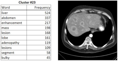 Automated labeling of CT images using deep learning. The image on the right was determined to fall into a similar cluster of images having the labels listed in the table on the left. Lead developers in NIH group: Xiaosong Wang and Le Lu.
Polyp detection is ready
Polyp detection is an area in which many groups have worked over the past 10 to 15 years, publishing more than 50 papers and developing highly accurate processing algorithms for detecting polyps. Polyp detection CAD was first commercialized in 2010, making this technique a reality, Summers said.
Organ volumetric analysis has been reported by multiple groups, for example, in automated liver and spleen and kidney volumetry. "The techniques have been shown to be robust in the presence of organomegaly and widespread metastases," Summers said. Unfortunately, these techniques are not in widespread use, although semiautomated volumetry of the liver and spleen is available.
Another technique presented by Liu et al in 2013 detects ovarian cancer metastasis, at least in perihepatic and perisplenic locations. There are no generalizable peritoneal detectors at this time, Summers said.
Renal lesions solved
Renal calculus detection and quantitation, also by Jianfei Liu and colleagues (SPIE, 2013), performs exceptionally well. The process identifies the kidney, smoothes the volume, and then detects calculi via maximally stable extremal region (MSER) feature extraction and support vector machine (SVM) classification. The system performs at over 90% sensitivity combined with an extremely low false-positive rate, making it a very useful application, Summers said.
Renal lesions can also be detected based on noncontrast CT colonography data, he said. Liu et al showed how exophytic lesions are detected by means of a bulge in the cortex of the kidney, and the same technique detects nonexophytic lesions in the setting of contrast-enhanced CT.
Automated pancreas shows promise
Pancreas detection and segmentation has recently taken off as well, according to Summers.
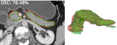 Automated segmentation of the pancreas. Red: manual annotation; green: automated segmentation. Lead developers: Holger Roth, PhD, and Le Lu, PhD.
"We placed a training dataset on the internet along with annotations, and a number of groups have been picking up on this and advancing the status of the field quite rapidly, so we're now getting accuracies in the low- to mid-80th percentile for pancreas segmentation," Summers said. "I think this will have applications for tumor detection and analysis and diabetes."
The NIH team has also made a dataset publicly available for lymphadenopathy detection, a field that's very important to the oncology community, he said. Despite big challenges, performance of the system by Lu et al continues to improve.
Then there is the spine, an area typically thought of as the domain of neuroradiology. But every abdominal CT contains a spine CT, which lends itself to analysis including osteoporosis scanning and lesion detection, Summers said. The group published two papers in Radiology regarding the detection of sclerotic and lytic vertebral body lesions and one paper in press in the Journal of Medical Imaging on mixed lesions. Another group showed that compression fractures are frequently overlooked by body radiologists on routine interpretation. The automated technique uses a height compass map that is similar to the one used in nuclear medicine.
Epidural masses can be a manifestation of metastatic disease in oncology patients, and the masses are frequently overlooked by radiologists if they don't pay special attention to the spinal canal during their assessment of abdominal CT, he said. Liu et al presented an automated technique for detecting them several years ago.
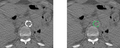 Left, original abdominal CT image. Right, automated segmentation of the calcified aortic plaque. Lead developer in NIH group: Dr. Jiamin Liu.
BMD with every abdomen scan
Bone mineral density (BMD) computed on the supine and prone scans of CT colonography data is reproducible within 10 mg/cc regardless of patient positioning, according to Summers.
Another NIH interest, muscle segmentation, has also been shown to be accurate and useful for assessing sarcopenia -- a risk factor for preoperative and oncology patients. Additionally, results with an automated muscle detector by Zhang et al were nearly identical to manual assessment, he said.
Many institutions are performing visceral fat assessments using semiautomated techniques, but a study by Summers' group showed the process can be fully automated, he said.
Vasculature as a road map
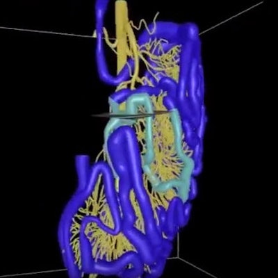
In the bowel, the mesenteric vasculature can be used to detect the small bowel on CT angiography, Summers said. A YouTube video showing the process can be viewed here.
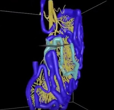 The mesenteric artery can be used to map the small bowel, an intricate organ that presents challenging imaging issues for clinicians looking for lesions, polyps, and tumors. The yellow lines represent vasculature, while the blue lines indicate the small bowel. Image courtesy of Dr. Tan Nguyen and Dr. Ronald Summers, PhD.
Finally, a new NIH study soon to be published shows accurate detection of colitis on CT scans in a fully automated way.
"We have a large population of patients on immunotherapy, and they not infrequently develop colitis," Summers said. The application "may be useful in certain classes of oncology patients."
"Fully automated assessment of the abdomen is advancing rapidly, with many problems tackled," he concluded. "New computer science tools we've heard about promise dramatic improvements, and I believe the quantitative radiologist's report of the future is close to reality."


