PET/MRI performs better than standard imaging for diagnosing cancer that has spread to the peritoneum from primary tumors in the abdominal cavity, according to research presented at RSNA 2021 in Chicago.
Researchers from Harvard Medical School compared imaging results between PET/MRI and PET/CT, CT, and MRI alone for diagnosing peritoneal carcinomatosis in patients with primary abdominal malignancies. They found PET/MRI offers significant advantages detecting cancer over standard approaches.
"PET/MRI provides a substantial increase in sensitivity for peritoneal involvement over [standard care imaging]," said radiology research fellow Dr. Felipe Furtado.
Peritoneal carcinomatosis refers to cancer that has spread to the lining surfaces of the peritoneum from a primary tumor in the abdominal cavity. Survival rates are low, as patients in the early stages often have few symptoms until the disease is fairly advanced. Current standard modalities are suboptimal and no studies to date have compared whether hybrid PET/MRI can improve diagnosis, according to Furtado.
"The detection of [peritoneal carcinomatosis] noninvasively by imaging is of compelling clinical interest in cancer staging and restaging," he said.
In the study presented at RSNA, Furtado and colleagues analyzed imaging of 164 patients (85 women) performed at Massachusetts General Hospital in Boston between April 2019 and March 2021. Patients were included if they had biopsy-confirmed primary abdominal or pelvic cancer and an FDG-PET/MRI scan within four weeks of imaging by a standard modality. Standard imaging consisted of whole-body contrast-enhanced (CE) PET/CT, abdominal CE-CT, or abdominal CE-MRI.
Two experts interpreted the scans, with a four-week interval between PET/MRI and standard imaging reads to reduce recall bias. Patients were considered positive if at least one lesion suspected of peritoneal carcinomatosis was detected on imaging.
Overall, PET/MRI detected 36 true-positive cases of peritoneal carcinomatosis compared with 20 true positives identified by standard imaging.
| Modalities compared in diagnosing peritoneal carcinomatosis | ||||
| PET/CT | CT | MRI | PET/MRI | |
| Sensitivity | 59% | 50% | 67% | 90% |
| Specificity | 96% | 94% | 100% | 92% |
| Accuracy | 88% | 88% | 95% | 91% |
"We can conclude that PET/MRI has a higher sensitivity when compared to standard of care imaging, while there was no significant difference between the standard of care imaging and PET/MRI specificity," Furtado said.
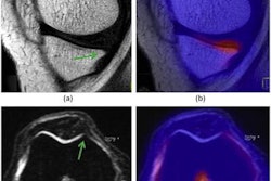
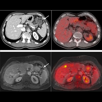
 A case of peritoneal thickening not seen on CT and also showing no FDG avidity. Enhancement of pulse-contrast MRI sequences detected peritoneal carcinomatosis, which was later confirmed in surgery. Image courtesy of Dr. Felipe Furtado.
Peritoneal carcinomatosis is not uncommon in gastrointestinal cancers, which account for one in three cancer deaths globally, and its detection on imaging is paramount to establish adequate patient management, Furtado said. Ultimately, this study shows that negative findings on PET/MRI can rule out peritoneal carcinomatosis with high confidence, he added.
"PET/MRI presents improved sensitivity for peritoneal carcinomatosis over the currently used modalities," he concluded.