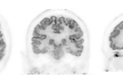
F-18 florbetaben PET scans for diagnosing Alzheimer’s disease can be reduced from 20 minutes to five minutes without losing accuracy, according to a recent study.
The finding is from an analysis of 307 PET scans from a memory clinic in South Korea and suggests the reduced time can overcome the need for rescans due to patient head movements, noted lead author Phuong Trinh, PhD, of Chonnam National University in Gwangju, and colleagues.
“Our findings suggest that shorter scan times are a viable and effective option for brain amyloid PET imaging in clinical settings,” the group wrote. The study was published November 21 in EJNMMI Research.
F-18 florbetaben PET (Neuraceq, Life Molecular Imaging) PET scans are used widely to visualize beta-amyloid deposits in the brain, a key marker used to diagnose Alzheimer’s disease. The scans typically involve a 20-minute acquisition, yet maintaining such prolonged scans can be challenging for elderly patients with memory impairment, the authors explained.
Moreover, within an enclosed space, most patients with psychiatric conditions often feel anxious or frightened by the buzzing and clicking sounds that occur during the scans, which can lead to head movements that compromise the qualitative and quantitative aspects of the images, they added.
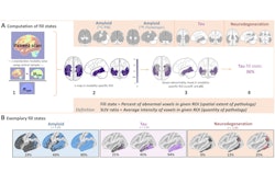
To investigate whether shorter scans can overcome these challenges, the group compared visual assessments among PET scans reconstructed after five, 10, 15, and 20 minutes. Nuclear medicine physicians validated and rated each scan as either amyloid-positive or negative. Further, the researchers compared PET metrics, namely total standardized uptake value ratio (SUVR) and Centiloid values, across five brain subregions: global, frontal, posterior cingulate-precuneus, lateral temporal, and parietal regions.
According to the findings, out of 307 cases, 199 were classified as positive for Alzheimer’s disease and 108 patients were negative. A comparison of images at five, 10, 15, and 20 minutes revealed that SUVR and Centiloid values gradually increased with prolonged scan times. The mean SUVR difference between the five- and 20-minute scans was 0.03 for the amyloid-positive and 0.01 for the amyloid-negative groups, while Centiloid differences were 4.6 and 2.38.
“We demonstrated that reducing the scanning duration from 20 to five minutes does not significantly affect the quantitative analysis of brain F-18 florbetaben PET images. Our results showed no significant difference in diagnostic confidence between the five-minute and 20-minute PET scan durations,” the researchers wrote.
Ultimately, while the five-minute scan may be suitable for the general population, certain subgroups, such as patients with higher BMI or obesity may require a tailored approach due to significant differences in the biodistribution of F-18 florbetaben in these patients, the researchers noted.
Also, a key limitation of shorter scan durations is the potential for reduced image resolution and clarity, which could introduce variability among nonexpert readers when identifying small amyloid lesions, the researchers added.
Nonetheless, the study supports the potential use of shorter scan durations as a viable diagnostic alternative, particularly among patients who experience discomfort or anxiety during longer scans, they suggested.
“This adaptation could improve patient compliance and expand the clinical utility of florbetaben PET imaging,” the group concluded.
The full study is available here.


