Advances in treatment-planning technology such as intensity-modulated radiation therapy (IMRT) are helping practitioners focus radiation on the tumor site and spare surrounding tissue. However, even this approach is not necessarily subtle enough for lung cancer. The condition of "healthy" lung tissue in patients with NSCLC is rarely uniform. An optimal radiotherapy treatment plan needs to identify which parts of a patient's lungs are working best, and ensure that these areas are not in the line of fire.
The idea is simple enough; however, finding the best way to mesh lung function data with IMRT is less straightforward. Researchers at the M. D. Anderson Cancer Center in Houston decided to examine two alternative options for mapping the quality of NSCLC patients' lung tissue: 4D CT and SPECT combined with standard CT (SPECT/CT).
In the first study, 4D CT performed at maximum exhalation and inhalation was used to generate ventilation images for 21 patients. Image data were imported into treatment-planning software to identify the 10% of lung volume in which ventilation was highest. The team members then designed IMRT plans to avoid this region and compared these with a baseline IMRT plan that took no account of the position of highly functioning lung (International Journal of Radiation Oncology, Biology, Physics, June 1, 2007, Vol. 68:2, 562-571).
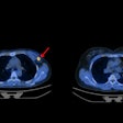
In a separate study, 16 patients underwent lung perfusion imaging with SPECT/CT prior to radiotherapy. As before, the imaging data were used to map areas of high function in the normal lung. IMRT plans created with and without input on lung function were then compared (IJROPB, August 1, 2007, Vol. 68:5, pp. 1349-1358).
Results from the two investigations were similar. In each case, the researchers concluded that the addition of functional data -- whether from 4D CT or SPECT/CT -- could help reduce the dose delivered to the healthiest parts of a patient's lungs.
"We want to demonstrate the potential benefit of using functional imaging, to show what we can achieve with it," stated radiation oncologist Dr. Thomas Guerrero, Ph.D., who participated in both studies.
So which one to choose? "Both of these investigations were just treatment-planning studies; we haven't used either technique in clinical practice yet," Guerrero said. SPECT-based perfusion imaging is a well-accepted technique, while 4D CT ventilation imaging is still relatively experimental. Guerrero has now acquired funding from the U.S. National Institutes of Health to validate the use of 4D CT-derived ventilation imaging in image-guided radiotherapy.
The choice of imaging technique may also hinge on economics as much as efficacy. NSCLC patients scheduled for radiotherapy at M. D. Anderson will have a 4D CT examination already as part of the treatment-planning process. So adding lung ventilation data would cost little in terms of time or money. In contrast, NSCLC patients do not normally undergo SPECT as part of their radiotherapy workup. Inclusion of SPECT-derived lung perfusion data would mean another examination for patients, which at M. D. Anderson translates into a $2,000 bill each time.
The optimum solution may not even be one of these two. U.K. researchers have also suggested that hyperpolarized helium-3 MRI could provide input on lung function in IMRT treatment planning. "That's an even more costly method," Guerrero noted.
By Paula Gould
Medicalphysicsweb contributing editor
October 24, 2007
Related Reading
US demonstrates cell death after chemo in experimental NHL study, September 13, 2007
Accelerated breast radiotherapy leads to less toxicity, radiation exposure, September 10, 2007
© IOP Publishing Limited. Republished with permission from medicalphysicsweb, a community Web site covering fundamental research and emerging technologies in medical imaging and radiation therapy.


