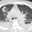
Respiratory Syncytial Virus
Clinical:
RSV is a common cause of bronchiolitis and pneumonia in children
and adults [1]. It is also the most common cause of lower
respiratory tract infection in infants [2]. RSV is the most common
viral pathogen in children hospitalized for community-acquired
pneumonia in the US [5]. Infection is common is the winter and
early spring [1] and children younger than 5 are more prone to
infection than older children [5]. Patients present with fever,
cough, dyspnea, and otalgia [1]. In immune competent adults the
infection usually produces rhinorrhea, pharyngitis, cough,
bronchitis, headache, fatigue, and fever [2]. In immunocompetent
patients, the course is usually self limited [1]. The infection
can be more severe and life-threatening in immune compromised
patients particularly in hematopoietic stem cell transplant
patients and those with hematologic malignancies [2]. RSV can be
an important illness in elderly adults with COPD accounting for
approximately 11% of hospitalizations for pneumonia [4].
X-ray:
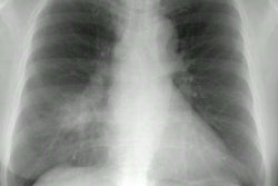
The majority of patients with RSV infection will have normal
radiographic findings [1]. in children, findings can include
central mpneumonia and peribornchitis [4]. In hospitalized adults
findings include consolidations and ground glass opacity- most
commonly unilateral and in a lower lung zone [4].
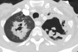
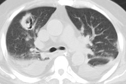
On CT, as RSV is known to predominantly cause the clinical
syndrome of bronchiolitis the infection is typically characterized
by evidence of airway inflammation with tree-in-bud opacities,
bronchial wall thickening, and peribronchiolar consolidaiton [3].
In immunocompromised patients findings include diffuse ground-glass opacification, consolidation, and tree-in-bud opacities [1].
REFERENCES:
(1) AJR 2005; Miller WT, Shah RM. Isolated diffuse ground-glass
opacity in thoracic CT: causes and clinical presentations. 184:
613-622
(2) Radiology 2011; Franquet T. Imaging of pulmonary viral
pneumonia. 260: 18-39
(3) AJR 2011; Miller WT, et al. CT of viral lower repiratory
tract infections in adults: comparison among viral organisms and
between viral and bacterial infections. 197: 1088-1095
(4) AJR 2014; Wong SSm, et al. Initial radiologic features as
outcome predictor of adult respiratory syncytial virus respiratory
tract infection. 203: 280-286
(5) Radiographics 2018; Koo HJ, et al. Radiographic and CT features pf viral pneumonia. 38: 719-739
