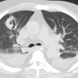
Influenza Pneumonia
- Clinical:
Influenza pneumonia is most commonly associated with type A and rarely type B organisms [1]. Influenza replicates in the respiratory epithelial cells and replication peaks approximately 48 hours after inoculation [1]. The early stages of disease often demonstrate tracheobronchitis and neutrophilic bronchopneumonia [1]. Secondary bacterial pneumonia can occur, particularly with S. pneumoniae [1].Avian flu is caused by H5N1 subtype of influenza type A and mortality is as high as 60% [1].
- X-ray:
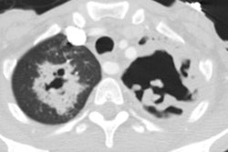
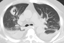
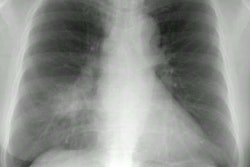
Radiographs show bilateral reticulonodular areas of opacity with or without focal areas of consolidation, usually in the lower lobes [1]. Poorly defined patchy or nodular areas of consolidation that become rapidly confluent (due to DAD) are seen frequently [1]. Pleural effusion is rare [1]. The presence of lobular consolidation suggests superimposed bacterial infection [1].REFERENCES:
(1) Radiographics 2018; Koo HJ, et al. Radiographic and CT features pf viral pneumonia. 38: 719-739
