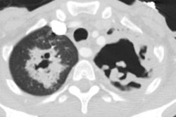
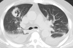
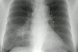
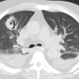
Haemophilus influenza:
Clinical:
H influenza is responsible for about 5-20% of community acquired pneumonias and is also an important cause of nosocomial infection [1].
X-ray:
CXR demonstrates patchy or segmental areas of consolidation [1]. Less commonly areas of non-segmental consolidation can be seen [1]. A reticular or reticulonodular interstitial pattern, by itself or in combination with air-space consolidation occurs in 15-30% of cases [1]. Cavitation occurs in 15% or less of cases [1]. Pleural effusion can be seen in up to 50% of cases, but empyema is uncommon [1].
REFERENCES:
(1) Radiol Clin N Am 2005; Tarver RD, et al. Radiology of community-acquired pneumonia. 43: 497-512
