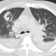
Lipoid Pneumonia:
Clinical:
Lipoid
pneumonia can be either exogenoous or
endogenous [4].
Exogenous lipoid pneumonia is caused by chronic aspiration or
inhalation of
mineral (paraffin, kerosene, or petroleum jelly), vegetable, or animal
oils
present in food, or oil based medications (such as nose drops). Once in
the
alveolar space, the oily substances are emulsified by lung lipase,
resulting in
a foreign body reaction. Predisposing conditions include neuromuscular
disorders
or structural abnormalities of the pharynx and/or esophagus that
predispose
them to aspiration [4]. Clinically patients with chronic lipoid
pneumonia are
frequently asymptomatic [4]. Symptomatic patients present with chronic
cough or
dyspnea, and less commonly fever, weight
loss, chest
pain, or hemoptysis [4]. Many patients are
elderly.
Lipoid pneumonia is a cause of chronic consolidation (other etiologies
to
consider for this finding include bronchoavleolar
cell carcinoma, tuberculous pneumonia, and
pseudolymphoma).
Acute exogenous lipoid pneumonia is uncommon and is typically caused by an episode of aspiration of a large quantity of a petroleum-based product [4]. It typically occurs in children (due to accidental poisoning), but can also occur in performers (fire-eaters) who use liquid hydrocarbons for flame blowing [4]. Patients present with cough, dyspnea, and low-grade fever [4].Radiographic opacities can be seen within 30 minutes of the episode of aspiration and will appear in most patients within 24 hours [4]. The opacities are typically ground glass or consolidative, bilateral, involve the middle or lower lobes, and are segmental or lobar in distribution [4]. Pneumatoceles can occur within 2-30 days and are more common in patients who have aspirated a large amount of mineral oils or petroleum-based products (such as can occur with fire-breathers) [4]. CT can reveal fat attenuation (-30 HU) in the areas of consolidation, but the associated inflammation often causes the fat to be less conspicuous [4]. The opacities resolve over 2 weeks to 8 months- resolution is generally complete, but mild scarring can occur [4].
Endogenous lipoid pneumonia results from lipid accumulation within intraalveolar macrophages in the setting of bronchial obstruction- "cholesterol pneumonia" [4]. It can also be seen in chronic pulmonary infection, pulmonary alveolar proteinosis, juvenile rheumatoid arthritis [5], or fat storage diseases [4]. Despite the presence of lipid, the consolidation does not appear of low attenuation on CT [4].
X-ray:
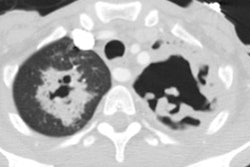
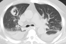
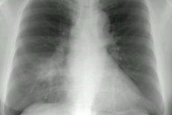
Radiographically, exogenous lipoid pneumonia is characterized by the
presence of lower lobe or right middle lobe [3] consolidations, mixed
alveolar
and interstitial opacities, and ill-defined or irregular mass-like
opacities
(due to chronic inflammation and fibrosis) [4]. On CT- the
consolidation can have fat
attenuation values (typically -30 to -80) and this will aid in
differentiation from other mass lesions. A "crazy-paving" pattern in
which well defined areas of ground-glass attenuation are superimposed
upon septal thickening has also been
described [3] (Ddx for this appearance
includes: Pulmonary alveolar proteinosis
and bronchoalveolar
cell carcinoma).
In
JRA, lipoid pneumonia shows multiple pulmonary nodules, mainly in a centrilobular distribution- which likely
represent
intra-alveolar and interstitial cholesterol granulomas
resulting from macrophage activation [5].
REFERENCES:
(1) AJR 1998; Franquet
T, et al.
The crazy-paving pattern in exogenous lipoid pneumonia: CT-pathologic
correlation.
170: 315-317
(2) Radiographics 2002; Gimenez A, et al. Unusual primary lung tumors: a radiologic-pathologic overview. 22: 601-619
(3) AJR 2008; Kim M, et al. MDCT evaluation of foreign bodies and liquid aspiration pneumonia in adults. 190: 907-915
(4) AJR 2010; Betancourt SL, et al. Lipoid pneumonia: spectrum of clinical and radiologic manifestations. 194: 103-109
(5) Radiographics 2011; Garcia-Pena P,
et al.
Thoracic findings of systemic diseases at high-resolution CT in
children. 31:
465-482
(6) AJR 2013; Nemec SF, et al. Lower lobe-predominant diseases of the lung. 200: 712-728
