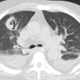
Viral Pneumonias (General):
Clinical:
Viruses can result in several forms of lower respiratory tract infection including tracheobronchitis, bronchiolitis, and pneumonia [1]. Adenovirus has the greatest effect in the terminal bronchioles [1].A viral etiology is the most common cause of pneumonia in children under the age of 5 years. Pathogens include: Respiratory syncytial virus (RSV), parainfluenza, adenovirus, influenza, enterovirus, and rhinovirus. Complications include: Subsequent bacterial pneumonia; Bronchiectasis; and Swyer-James Syndrome. The infection primarily affects the AIRWAYS (not the alveoli) therefore will see changes secondary to airblock.
In adults viral pneumonias are usually gradual in onset, have
associated
non-pulmonary symptoms, and typically only produce mild elevations
in
the
white blood cell count. RSV and parainfluenza virus typically
produce
only
upper airway infections or bronchitis (characterized by evidence
of
airway inflammation with tree-in-bud
opacities, bronchial wall thickening, and peribronchiolar
consolidaiton) [2]. Rhinovirus predominantly
involves
only the upper respiratory tract. Adenovirus infection typically
prresents as a multifocal pneumonia pattern with areas of
ground-glass
opacity or consolidation without airway abnormalities [2].
Influenza A is the most important respiratory viral illness with
more than 35,000 deaths and 200,000 hospitalizations annually [1].
Symptoms include rapid onset of high fever, myalgias, headache,
lethargy, sore throat, and cough [1].
Parainfluenza virus has been noted to cause pulmonary infection
particularly in patients who have received organ transplants [3].
It has also been reported as one of the more common viruses
associated with exacerbations of asthma in adults (along with RSV
and influenza virus) [3]. It commonly causes not only rhinitis and
sinusitis, but also lower tract disease including bronchitis,
bronchiolitis, and pneumonia [3]. On imaging, it often shows an
airway-centric disease pattern with tree-in-bud opacities,
bronchial wall thicekning, ground-glass opacities, and
peri-bronchiolar patchy consolidation [3].
Avian flu is caused by the H5N1 subtype of influenza A virus and
approximately 90% of infections have occurred in patients 40 years
old
or younger [1]. The fatality rate exceeds 60% [1].
Measles virus is a highly contagious infection with an incubation
time of almost 2 weeks [1]. It can result in a fatal pneumonia in
immunocompromised and debilitated patients [1].
X-ray:
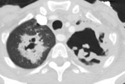
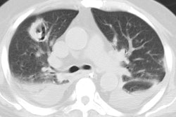
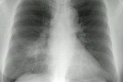
In children, the CXR characteristically demonstrates HYPERINFLATION with a reticular or airspace pattern. The infection is more commonly bilateral as opposed to lobar. Other findings include perihilar linear densities with loss of hilar and vascular sharpness, patchy atelectasis, and bronchial wall thickening (peribronchial cuffing). Adenopathy (3%) and effusion are uncommon.In adults, viral infections commonly produce a diffuse, fine reticular pattern. Bronchial wall thickening is also common. Severe infection can result in air space opacities- typically patchy in distribution. Patients that develop a pneumonia secondary to an influenza infection demonstrate a rapidly progressive bilateral pneumonia or patchy bronchopneumonia. Superinfection with Staphylococcus should be suspected if cavitation develops.
On HRCT, bronchial wall thickening, septal prominence, patchy
areas
of heterogeneous/ground glass attenuation, centrilobular ground
glass
nodules, airspace consolidation, and/or small branching nodular
opacities
in the lung periphery (tree-in-bud- indicative of bronchiolar
impaction)
can all be seen [1].
REFERENCES:
(1) Radiology 2011; Franquet T. Imaging of pulmonary viral
pneumonia.
260: 18-39
(2) AJR 2011; Miller WT, et al. CT of viral lower repiratory
tract
infections in adults: comparison among viral organisms and between
viral and bacterial infections. 197: 1088-1095
(3) AJR 2013; Herbst T, et al. The CT appearance of lower
respiratory infection due to parainfluenza virus in adults. 201:
550-554
