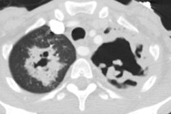
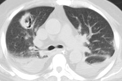
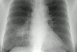
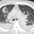
PCP with pneumothorax:
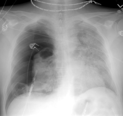
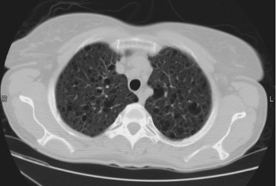
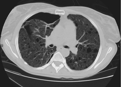
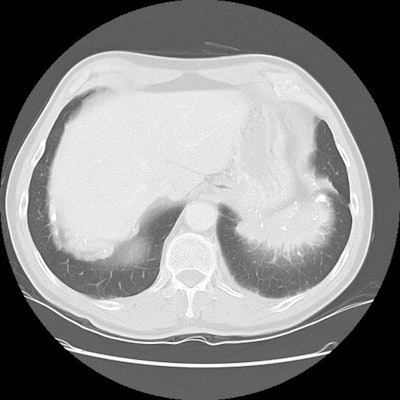
This is another example of pneumocytis pneumonia in a patient with cystic changes and a penumothorax. Extensive air space consolidation is seen in the left lung. CT (below) demonstrated the large right pneumothorax and the presence of areas of ground-glass attenuation. Interlobular reticulation within the areas of ground-glass reflects interstitial and interlobular septal infiltration by mononuclear cells and edema. This is often the predominant finding during the sub-acute phase of the illness. Thin walled cystic spaces are seen bilaterally. A thick walled cyst with an air-fluid layer is seen within the left lung.
(Click CXR to enlarge)
