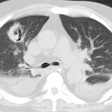
Accuracy of CT in the diagnosis of allergic bronchopulmonary aspergillosis in asthmatic patients.
Ward S, Heyneman L, Lee MJ, Leung AN, Hansell DM, Muller NL
OBJECTIVE: The purpose of this study was to assess the accuracy of high-resolution CT in the diagnosis of allergic bronchopulmonary aspergillosis in asthmatic patients. MATERIALS AND METHODS: The high-resolution CT scans of 44 asthmatic patients with allergic bronchopulmonary aspergillosis and 38 asthmatic patients without allergic bronchopulmonary aspergillosis were analyzed retrospectively and randomly by two independent observers for these features: bronchial wall thickening, bronchiectasis, centrilobular nodules, mucoid impaction, mosaic perfusion, atelectasis, and consolidation. Each observer made a final diagnosis with a stated degree of confidence. The results are expressed as the average number of observations by the two observers. RESULTS: Findings seen more commonly in patients with allergic bronchopulmonary aspergillosis than in patients with asthma alone included bronchiectasis, centrilobular nodules, and mucoid impaction (p < .01, chi-square test). Bronchiectasis was present in 42 (95%) of 44 patients with allergic bronchopulmonary aspergillosis, centrilobular nodules in 41 (93%), and mucoid impaction in 29.5 (67%) (average of two observers). In the asthmatic control group, bronchiectasis was detected in 11 (29%) of 38 patients, centrilobular nodules in 10.5 (28%), and mucoid impaction in 4%. Bronchiectasis was seen in 184 (70%) of 264 lobes of patients with allergic bronchopulmonary aspergillosis compared with 19.5 (9%) of 228 lobes in asthmatic controls (p < .001, chi-square test). CONCLUSION: In asthmatic patients, bronchiectasis affecting three or more lobes, centrilobular nodules, and mucoid impaction are findings on high-resolution CT that are highly suggestive of allergic bronchopulmonary aspergillosis.
PMID: 10511153, UI: 99439171
