Tularemia Pneumonia: Francisella tularensis
Clinical:
Francisella tularensis is a highly infectious gram negative coccobacillus which can remain viable in the environment for 3 to 4 months at low temperatures, in mud, water, and decaying animal carcasses. The organism does not grow on routine lab media, however, and requires cysteine enriched blood agar for growth. The most important reservoirs and vectors are ticks, hares, and rabbits. Hunters may acquire the disease while skinning wild animal meat. Following a bite or cut, a skin ulcer will develop. From the skin, the organism spreads to the lymphatics where it produces painful and sometimes suppurating regional adenopathy. This is followed by hematogenous spread to other organs. Six forms of tularemia have been described- ulceroglandular (70-80% of cases), glandular, oculoglandular, oropharnygeal, typhoidal (associated with a 30-60% mortality), and pleuropulmonary.Pleuropulmonary infection can occur secondary to bacteremic spread or from inhalation of infectious aerosols. The onset of symptoms following inhalational infection is usually abrupt with high fever, chills, dyspnea, non-productive cough, and sweating. When occurring as a complication of ulceroglandular or glandular infection, the signs and symptoms may be mild and non-specific and last a few days to weeks. In up to 25-30% of these patients, the CXR may demonstrate extensive infiltrates, but physical findings may be minor. Streptomycin is the treatment of choice.
X-ray:
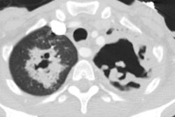
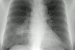
Secondary pneumonias usually present as bilateral lower lobe infiltrates with exudative pleural effusions in 60-80% of cases (usually bilateral). Other findings may include miliary infiltrates, lobar consolidation, hilar adenopathy without infiltrate, or mediastinal adenopathy.Images:
Case 1: Tularemia following skinning wild gameREFERENCES:
(1) Seminars in Respiratory Infections 1997; Tularemia pneumonia. 12 (1): 61-67


