Radiology 1997 Jul;204(1):171-175
Mycobacterium kansasii pulmonary infection in patients with AIDS: spectrum of chest radiographic findings.
Fishman JE, Schwartz DS, Sais GJ
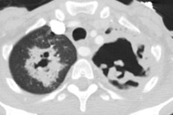
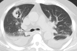
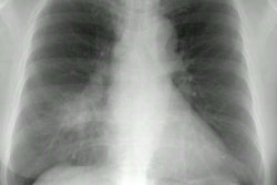
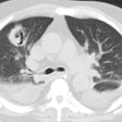
PURPOSE: To determine the chest radiographic findings and clinical manifestations of Mycobacterium kansasii pulmonary infection in patients with acquired immunodeficiency syndrome (AIDS). MATERIALS AND METHODS: Criteria for diagnosis included two or more positive cultures from respiratory sources, pulmonary symptoms or fever, and no other identifiable cause of pulmonary disease. Chest radiographs at initial examination and follow-up were evaluated for parenchymal opacities, cavitation, adenopathy, and pleural effusions. Medical records were reviewed for clinical signs and symptoms, CD4 cell count, presence of additional pathogens, and response to antimycobacterial therapy. RESULTS: Of 96 patients, 16 (17%) satisfied all criteria for M kansasii pulmonary infection. The mean CD4 cell count was 24/mm3. Twelve patients (75%) demonstrated alveolar opacities, only three (19%) of which were cavitary. Interstitial opacities (6%) and pleural effusions (12%) were uncommon. Four (25%) patients had thoracic lymphadenopathy, which was the only positive radiographic finding in two patients. Fourteen patients were treated for M kansasii, and 10 (71%) showed clinical and radiographic improvement. CONCLUSION: Patients with AIDS and pulmonary M kansasii frequently demonstrate focal alveolar opacities. Symptomatic patients with pulmonary nontuberculous mycobacteria should be presumptively treated for pulmonary M kansasii until final culture results are available.
PMID: 9205241, MUID: 97349237
