Paragonimiasis:
Clinical:
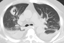
Paragonimiasis westermani (the lung fluke) infection is common in Southeast Asia and the Far East. The organism is ingested with raw crab or crayfish [3]. The encyst penetrates the intestinal wall and migrates through the peritoneal cavity. Most penetrate the diaphragm and pleura and mature to adult flukes in the lungs. In the lungs the organisms localize near small bronchioles, incite inflammation, and the formation of a fibrous capsule in which adult worms and ova are found. The eggs penetrate the bronchioles and exit the host- by either being coughed up or swallowed and passed in the stool [3]. Alternatively, the eggs may end up in the pleural space- a dead end for reproduction- where they incite an intense inflammatory host response [3]. Patients present with symptoms of a cough productive of brownish sputum similar to chronic bronchitis. Hemoptysis can occur. Treatment is with praziquantel and therapy may result in worsening of the radiographic findings.P. kellicotti is the organizm that causes North American paragonimiasis [3]. Infection can result in pleural and pericardial effusion, internal mammary and cardiophrenic angle adenopathy, and peripheral lung nodules with surrounding ground-glass opacity [3]. A linear track extending from the nodule to the pleura is suspected to correspond to the worms burrow tunnel [3].
X-ray:
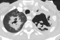
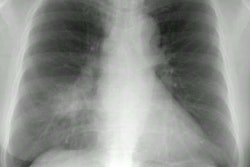
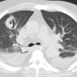
CXR demonstrates migratory infiltrates, but after 1 to 2 months the lesions become stable nodules or cysts predominantly in the lower lobes. Pleural effusions can be seen.CT findings include poorly marginated peripheral or subpleural nodules measuring approximately 2cm (up to 74% of cases) with surrounding ground-glass and a streaky opacity connecting the nodule to the pleural surface [3], pleural effusion, hydropneumothorax, airspace consolidation, and thin walled cysts [1,2].
The focal lesions can be hot on FDG PET imaging [1].
REFERENCES:
1. AJR 2005; Kim TS, et al. Pleuropulmonary paragonimiasis: CT findings in 31 patients. 185: 616-621
(2) Radiographics 2007; Jeong YJ, et al. Eosinophilic lung diseases:
a clinical, radiologic, and pathologic overview. 27: 617-639
(3) AJR 2012; Henry TS, et al. Chest CT features of North American
paragonimiasis. 198: 1076-1083
