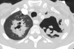
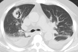
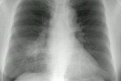
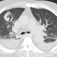
Pneumatocele:
Clinical:
A pneumatocele is an acquired lung cyst. They are felt to
most likely represent a subpleural collection of air
formed by dissection of air from ruptured bronchioles or alveoli through the interstitium of the lung. Pneumaoceles
are commonly seen in infants/small children due to obstruction of smaller
bronchi with resultant air trapping.
They
occur secondary to:
1- A
preceding supporative pulmonary infection: Typically
occur in infants and young children- with 70% of cases in children under the
age of 3 years [1]. Post-infectious pneumatoceles are
rare in adults [1]. They most frequently complicate staphylococcal pneumonia,
but can also be seen less commonly following pneumococcus,
E. coli, Klebsiella, or haemophilus
influenza pneumonia [1]. The pneumatocele typically
appears within 1 week of onset of the infection and most spontaneously resolve
over weeks (usually within 6 weeks) to months [1].
2- Trauma- Traumatic pneumatoceles appear as round, or oval areas of radiolucency with thin or imperceptible walls. They are the result of a lung laceration injury due to deceleration injury- air/gas becomes trapped within area of parenchymal laceration [1]. They can be identified as early as one hour following the injury, or may appear later as an area of parenchymal contusion clears. Post traumatic pneumatocele may be solitary or multiple, generally spare the lung apices, and often resolve within 3 weeks [1], although other aurthors indicate that post-traumatic pneumatoceles clear more slowly than psot-infectious ones.
3-
Hydrocarbon inhalation
A pneumatocele is completely air filled, unless superinfected and they generally resolve spontaneously, but
they may enlarge.
X-ray:
On
imaging, a pneumatocele appears as a well-defined
cystic structure with a thin wall [1].
REFERENCES:
(1) Radiographics 2011; Dillman JR, et al. Expanding upon the unilateral hyperlucent hemithorax in children. 31: 723-741
