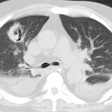
Strongyloides:
Clinical:
Infection with Strongyloides stercolis (a common nematode) occurs throughout the world and is associated with poor sanitation. The organism enters the body via the hands or feet and migrates via the lymphatics and venous system through the right side of the heart to the lungs. The larvae penetrate the capillary walls into the alveoli, ascend the tracheobronchial tree, are swallowed, and mature in the upper part of the small intestine. Patients present with blood eosinophilia, bloody diarrhea, and bronchospasm or pneumonia-like symptoms. It may remain in the lung and develop into mature worms that reproduce locally. Treatment is with Thiabendazole.
In patients with cell-mediated immunity defects, Strongyloides hyperinfection syndrome can develop with diffuse pulmonary opaciities, gram-negative sepsis, respiratory failure, and a high mortality rate [1].
X-ray:
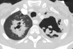
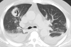
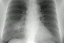
The chest radiograph can be normal. During migration through the lungs, a fine miliary nodular or diffuse reticular interstitial opacities can be seen. With heavier infestations confluent areas of opacification may be present. Rarely, cavitary lesions or pleural effusions can be seen.
REFERENCES:
(1) Radiographics 2007; Jeong YJ, et al. Eosinophilic lung diseases: a clinical, radiologic, and pathologic overview. 27: 617-639
