AJR Am J Roentgenol 1997 Sep;169(3):649-653. Tuberculosis of the central airways: CT findings of active and fibrotic disease.
Moon WK, Im JG, Yeon KM, Han MC
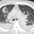
OBJECTIVE: Management of patients with central airways tuberculosis differs according to the activity of the disease. The purpose of this study was to analyze CT findings of active and fibrotic disease in patients with central airways tuberculosis. MATERIALS AND METHODS: According to bronchoscopic findings and biopsy results, 41 patients with tuberculosis of the trachea and main bronchi were categorized as having active disease (n = 30) or fibrotic disease (n = 11). Follow-up CT scans were obtained after antituberculous therapy in 11 patients with active disease and two patients with fibrotic disease. All CT scans were retrospectively analyzed with particular attention to the locations of airway lesions, patterns of luminal narrowing, wall thickening of diseased airways, and presence of abnormal adjacent lymph nodes. RESULTS: Active disease in 30 patients involved the trachea (n = 20), the right main bronchus (n = 14), or the left main bronchus (n = 13). Seventeen patients had multiple lesions. On CT scans, these airways showed irregular (n = 24) or smooth (n = 4) narrowing in 28 patients: minimal (n = 5) or marked (n = 18) wall thickening with contrast enhancement in 23 patients: and obstruction with peribronchial cuffing in nine patients. Enlarged mediastinal lymph nodes were seen in 26 patients. Fibrotic disease in 11 patients involved the trachea (n = 6), the right main bronchus (n = 2), or the left main bronchus (n = 9). Six patients had multiple lesions. On CT scans, the airways showed smooth (n = 7) or irregular (n = 2) narrowing without (n = 5) or with minimal (n = 4) wall thickening in nine patients and obstruction without peribronchial cuffing in four patients. On follow-up CT scans, the findings for the airway lesions were almost normal in nine patients who had had initial active disease. However, the findings for airway narrowing did not change in two patients with fibrotic disease after 6 months of follow-up. CONCLUSION: Principal CT findings in our patients depended on disease stage. Central airways narrowing was seen in both active and fibrotic stages. However, in patients with active disease, CT scans showed irregular and thick-walled airways, a pattern that was reversible, whereas patients with fibrotic disease generally had smooth narrowing of airways and minimal wall thickening, a pattern that was not reversible during the follow-up period.
PMID: 9275870, MUID: 97421753
