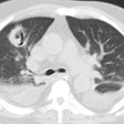
Round Pneumonia:
View cases of round pneumonia
Clinical:
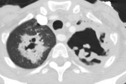
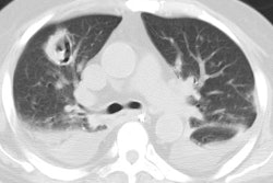
Round pneumonia is classically described in children and is almost always bacterial- especially pneumococcal. It is most commonly located in the posterior lower lobe.
Round pneumonia may also be seen in adults (in as many as 10% of cases of pneumonia) [1]. It can be associated with a compromised immune response and may be fungal. The clnical symptoms in non-immune compromised patients may be mild and mimick a viral syndrome or bronchitis. A trial of antibiotics followed by a repeat chest radiograph in 2 to 3 weeks is probably a reasonable course of action in any patient with a solitary pulmonary nodule [1]. Any lesion that persists, should then be further evaluated to exclude malignancy [1].
X-ray:
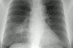
On CXR it appears as a sharply defined, rounded, soft tissue density. With antibiotic therapy it becomes poorly defined, then rapidly clears. Air bronchograms are seen in only about 20% of cases. Other findings that can be seen in association with a round pneumonia include adjacent pleural thickening and satellite nodules (best appreciated on CT).
REFERENCES:
(1) AJR 1998; Wagner AL, et al. Radiologic manifestations of round pneumonia in adults. 170: 723-726
