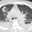
Coccidiomycosis:
Clinical:
Coccidiomycosis (valley fever) is the second most common endemic fungal infection encountered in the United States. The fungus is found in soil and occurs primarily in the southwest U.S. and Northern Mexico [4]. The two principal species that cause human infection are C. immitis (found in the San Joaquin valley of California) and C. posadasii (found outside of California) [4]. The hyphal form proliferates during the rainy season and dies in the dry period, releasing highly infectious spores (anthroconida) [3]. The infection is acquired by inhalation of arthrospores contained in dust. After inhalation, the spores swell into spherules in the small airways and reproduce through cleavage [23]. The mature spherules rupture and release endospores causing inflammation [3]. The incidence of disease is seasonal, with most cases occurring in the late summer, fall, and winter [4].Primary Pulmonary Infection:
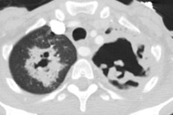
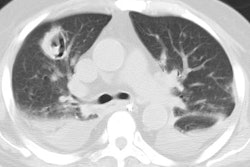
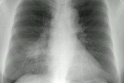
The primary infection is usually benign and asymptomatic in 60% of cases. In symptomatic patients a dry cough may be associated with fever, arthralgias, headache, or night sweats producing an influenza-like illness. Most cases occur in adulthood [23]. Individual risk factors include extreme ages of life, smoking, male sex, pregnancy, hematologic malignancy, underlying lung disease, and diabetes [4]. Cutaneous lesions such as erythema nodosum or erythema multiforme can occur in up to 25% of white women and 4% of white men [3]. The primary infection usually resolves over 2 to 6 weeks in 85-95%.Patchy parenchymal consolidations are found in 75% of symptomatic patients and are mostly unilateral with a perihilar and basilar predominance [3]. The infiltrates have a propensity to develop in one site as another site clears ("phantom infiltrates"). Well defined lung nodules are another manifestation of the infection and are found in about 20% of cases. Calcification in a coccidiomyoma is much less frequent than seen in a tuberculoma or a histoplasmoma. Cavitary lesions can be seen in 2-8% of hospitalized patients with acute infection [3] and have classically they have characteristic thin walls ("grape-skin" like). About 90% of the cavities are solitary, within the upper lobe, and lack an air-fluid level.
Hilar adenopathy is noted in 20-40% of cases of acute infection [4] and may persist for months to years (particularly mediastinal adenopathy). Rarely, adenopathy will be the only imaging finding (8% of cases) [4]. Small, unilateral transdative or exudative pleural effusions are found in about 5-20% of cases [3,4].
Chronic Pulmonary Infection:
When the clinical symptoms and radiologic findings persist for more than 6 weeks, the patient is considered to have a chronic infection (affecting 5-15% of patients [4]), however, only 25% of patients with chronic changes have a history suggestive of antecedent acute coccidiomycosis. Manifestations of chronic disease include air space consolidation, nodules, cavities, pleural disease, bronchiectasis, and fibrosis.Chronic Progressive Coccidioidal Pneumonia:
Chronic progressive infection develops in 1-5% of patients and is diagnosed when clinical symptoms or imaging abnormalities persist beyond 6 weeks [3]. Findings include residual nodule (most common), persistent pneumonia, or persistent pleural effusion [3]. Apical fibronodular and cavitary disease associated with volume loss is another manifestation and patients can be asymptomatic [3]..Disseminated Cocciciomycosis:
Disseminated disease occurs in less than 1% of cases overall and up to 11% of hospitalized patients [3]. Disseminated disease occurs more commonly in blacks (12x's higher than whites), Hispanics, and orientals (Filipino). There is an increased chance for dissemination in infants and the elderly. Patients with diabetes, on dialysis, and the immune compromised (HIV, steroids, or immuno-suppressive medication) are also at increased risk [4]. Hematogenous dissemination commonly involves the lung, skin, musculoskeletal system, and the meninges. Disseminated disease is fatal in 50% of cases without treatment.Disseminated infection is characterized radiographically by the presence of ill-defined miliary nodules (due to hematogenous spread with a random distribution [4]), and associated hilar and mediastinal adenopathy is nearly always present [4].
REFERENCES:
(1) Semin Roentgenol 1996; Thoracic coccidioidomycosis. 31(1), 28-44 (No abstract available)
(2) J Thorac Imag 1999; Conces DJ. Endemic fungal pneumonia in
immunocompromised patients. 14: 1-8
(3) Radiographics 2014; Jude CM, et al. Pulmonary
coccidiomycosis: pictorial review of chest radiographic and CT
findings. 34: 912-925
(4) Radiographics 2021; Kunin JR, et al. Thoracic endemic fungi in the United States: importance of patient location. 41: 380-398
