Nocardia:
Clinical:
Nocardia is a gram-positive and weakly acid fast aerobic organism
found in soil and
decaying vegetation. Most cases of pulmonary infection are caused by N.
asteroides (N. asteroides accounts for approximately 85% of all
nocardial infections and most pulmonary infections [4]).
Infection is common in immune compromised hosts (particularly those
with impaired cell mediated immunity [4]), diabetics, and patients
receiving high
dose corticosteroids. Other risk factors for infection include:
Immunosuppression
(following organ transplant, esp. cardiac transplantation), alveolar
proteinosis, HIV infection [4], and
lymphoreticular malignancy (leukemia, lymphoma). The reported incidence
of nocardial infection in solid organ transplant patients varies from
0.7-3.5% [4].
Patients generally present with cough, fever, dyspnea, and constitutional symptoms, but a wide range of clinical presentations can be seen. Nocardia can spread hematogenously to extrapulmonary locations- most frequently to the CNS with brain abscess, but also to the skin and joints [4]. Approximately one-third of infected immunocompromised patients will have disseminated disease [4]. Diagnosis is often difficult owing to the slow growth of the organism. Treatment with sulfonimides is usually effective.
X-ray:
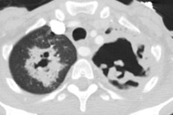
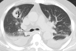
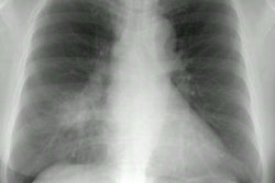
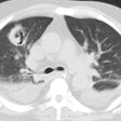
The infection typically begins as a localized consolidation which
progresses to
cavitation. The consolidation can appear mass-like. Chest wall
extension with rib
destruction and sinus tract formation can be seen. Multiple pulmonary
nodules which can
cavitate (33%) are another common manifestation of infection,
particularly in HIV patients
[2]. Pleural effusion occurs in approximately 50% of cases and
adenopathy can also be
identified. However, other authors state that mediastinal and hilar
adenopathy are not usual features of pulmonary nocardiosis [4].
REFERENCES:
(1) J Comput Assist Tomogr 1995; 19 (1): 52-55
(2) J Comput Assist Tomogr 1995; 19 (5): 726-732
(3) J Thorac Imaging 1998; Conces DJ.
Bacterial pneumonia in
immunocompromised patients. 13: 261-270
(4) AJR 2011; Kanne JP, et al. CT findings of pulmonary Nocardiosis.
197: W266-W272
