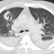
Actinomycosis:
Clinical:
Actinomyces israelii is a pleomorphic branching filamentous bacterium that is part of the normal oral flora [2]. Actinomycosis infection most commonly involves the cervicofacial region which represents more than 50% of cases (typically the result of poor dental hygiene, recent dental extraction, and dental caries) [3]. Pulmonary infection is frequently due to poor oral hygiene and disorders which predispose to aspiration (such as alcoholism) [2,3]. Clinical manifestations are productive cough, low-grade fever, and blood-tinged sputum [2]. Penicillin G is the drug of choice for treatment and the therapy often requires a prolonged course of antibiotics [3]. The prognosis is generally good with appropriate antibiotic therapy [2]. The organisms produce sulfur granules (which may be found in the sputum) and may directly invade chest wall or mediastinum (an important finding in making the diagnosis).X-ray:
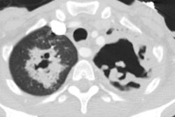
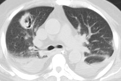
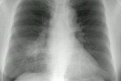
The infection begins as a local consolidation which may go on to cavitate. A unique feature of the infection is it's tendency to invade the chest wall producing sinus tracts. An associated pleural involvement (pleural thickening, pleural effusion, or empyema) is common (more than 50% of cases) [3]. On CT, segmental air-space consolidation with low attenuation regions, peripheral enhancement, and adjacent pleural thickening are commonly seen. Other findings on CT include hilar or mediastinal adenopathy, and pleural effusion [2]. The infection can sometimes cross an adjacent interlobular fissure or involve the chest wall [2].REFERENCES:
(1) Radiology 1998; Cheon JE, et al.
Thoracic
actinomycosis: CT findings. 209: 229-233
(2) AJR 2006; Kim TS, et al. Thoracic actinomyocosis: CT features
with histopathologic correlation. 186: 225-231
(3) Radiographics 2014; Hei SH, et al. Imaging of actinomycosis
in various organs: a comprehensive review. 34: 19-33
