Coxiella burnetii pneumonia: Q Fever
View images of Q fever
Clinical:
Q fever is a zoonosis caused by Coxiella burnetii - an
obligate intracellular bacteria
living in the phagosomes of
the host cells. It can be found in the urine, feces, and milk of
infected animals
(especially cattle, sheep, and goats). C. burnetti can survive for
long
periods of time in
the environment. Humans acquire the infection from inhaled dust or
by
ingesting
unpasteurized milk or cheese. Farmers and stock breeders are at
high
risk becasue of
contact with livestock. Clinical findings include prolonged fever,
pneumonia,
granulomatous hepatitis, and meningoencephalitis. Treatment is
with
tetracycline.
Chronic Q fever can develop in 1-5% of infected patients and it's
manifestations include endocarditis, infected aneurysms, or
infected vascular prostheses [3].
X-ray:
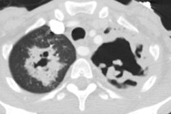
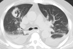
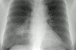
The most common radiographic finding is unilateral segmental or lobar consolidations. The consolidation can appear mass-like. Pleural effusion can be found in about 15% of cases. The most common finding on CT scan is multilobar air space consolidation [2].
REFERENCES:
(1) Radiology 1999; Gikas A, et al. Q fever pneumonia: Apperance
on
chest radiographs
(2) Radiology 2000; Voloudaki AE, et al. Q fever pneumonia: CT
findings. 215: 880-883
(3) J Nucl Med 2018; Kouijzer IJE, et al. The value of 18F-FDG
PET/CT in diagnosis and during follow-up in 273 patients with
chronic Q fever. 59: 127-133


