Distinguishing hantavirus pulmonary syndrome from acute respiratory distress syndrome by chest radiography: are there different radiographic manifestations of increased alveolar permeability?
Ketai LH, Kelsey CA, Jordan K, Levin DL, Sullivan LM, Williamson MR,
Wiest PW, Sell JJ
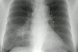
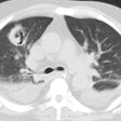
Hantavirus infection may cause diffuse air space disease, termed hantavirus pulmonary syndrome (HPS). The authors sought to determine if chest radiographs could differentiate HPS from typical acute respiratory distress syndrome (ARDS). The authors identified patients with either HPS (n = 11) or acute ARDS (n = 32) and selected the earliest chest radiograph showing diffuse airspace disease, and a chest radiograph taken 24 to 48 hours previously. Thoracic and general radiologists first viewed the chest radiograph showing diffuse air space disease, and ranked the likelihood that each case represented HPS versus ARDS. Afterward, readers viewed earlier chest radiographs and rescored each case. Receiver operating characteristic (ROC) curves from both scoring sessions were generated. The mean areas under the ROC curves for the entire group was 0.83 +/- 0.12 initially, and improved to 0.87 +/- 0.09 (p < 0.05) after viewing prior chest radiographs. Receiver operating characteristic curves of thoracic radiologists described greater areas than those of general radiologists both before and after viewing prior chest radiographs; 0.95 +/- 0.01 versus 0.78 +/- 0.08 (p < 0.05) and 96 +/- 0.02 versus 0.80 +/- 0.05 (p < 0.05). The mean sensitivity and specificity of chest radiograph interpretation for HPS was 86 +/- 13% and 74 +/- 11%, respectively. Chest radiographs can differentiate HPS from ARDS. Accuracy is improved by the use of serial radiographs and more highly trained readers. The chest radiograph findings may represent differences in the extent of alveolar epithelial damage seen in HPS and ARDS.
