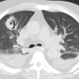
Old Histoplasmosis Infection with a Mediastinal Granuloma:
The patient was an active duty troop who presented for evaluation of URI symptoms which were probably unrelated to the radiographic findings. The patient was skin test positive for Histoplasmosis.
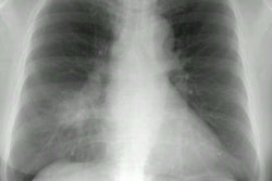
The CXR demonstrates a large right paratracheal mass. A more subtle finding is a calcified nodule within the left lower lung. (Click on small image to view larger radiograph)

The patient had a CT performed that demonstrated the paratracheal mass to be densely calcified (a finding not readily appreciable on the plain film). The calcified mass extended in to pretracheal region. The superior vena cava is displaced but there is no mediasitnal fibrotic reaction evident to suggest fibrosing mediastinitis. The calcified nodule was also again identified. (Click on small image to view larger radiograph)


