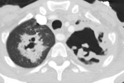
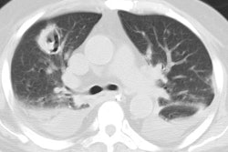
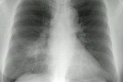
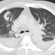
CT of fibrosing mediastinitis: findings and their utility.
Weinstein JB, Aronberg DJ, Sagel SS
The computed tomographic (CT) manifestations of fibrosing mediastinitis were assessed in seven patients with pathologically proven disease. Computed tomography had been done to evaluate further a mediastinal or hilar mass seen on the conventional chest radiograph or to define extent of disease preoperatively. Findings included a mediastinal or hilar mass (7/7), calcifications of the central mass or in associated lymph nodes (6/7), tracheobronchial narrowing (5/7), and pulmonary infiltrates (4/7). In six of the seven patients, CT demonstrated masses or calcifications that were not evident with conventional radiography. The CT findings often were sufficient to suggest or corroborate the diagnosis of fibrosing mediastinitis, and the extent of the disease process was well depicted. In selected patients the CT findings may be sufficient to exclude the need for diagnostic tissue sampling.
