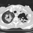
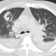
Fibrosing mediastinitis:
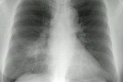
The patient had known fibrosing mediastinitis and presented for further evaluation after a nuclear medicine perfusion scan revealed absent perfusion to the left upper lobe. This case is similar to the previous one in that there has been complete obliteration of the left upper and lower pulmonary veins. This results in obstruction to pulmonary outflow with resultant decreased perfusion. See discussion below- click images to enlarge
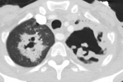
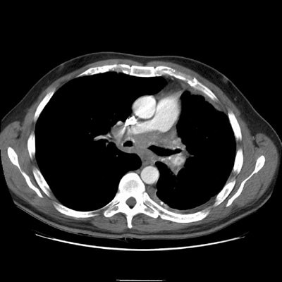
CT reveals an infiltrative mediastinal soft tissue abnormality encasing and narrowing the trachea.
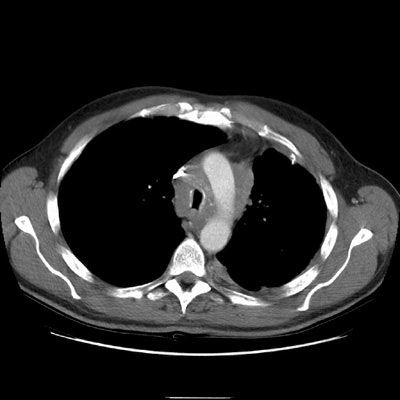
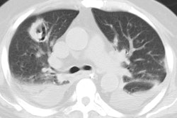
The right upper lobe pulmonary vein can be seen just lateral and slightly posterior to the SVC. It is markedly narrowed and distorted by the mediastinal infiltrative soft tissue abnormality. No left upper lobe pulmonary vein is identified. The left pulmonary artery structures are narrowed and irregular.
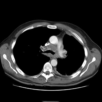
Again note abnormality involving the left pulmonary artery and airway narrowing.
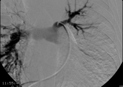
A pulmonary arteriogram revealed no left upper lobe perfusion. The right upper lobe pulmonary artery is narrowed (black arrow)
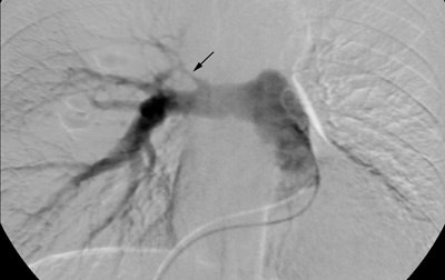
Selective left pulmonary arteriogram revealed the vessel to be patent, although irregular and narrowed. A delayed run was not performed, but the findings were felt to be consistent with pulmonary venous obstruction based upon the CT scan findings.
