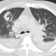
Cryptococcus:
Clinical:
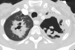
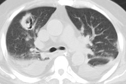
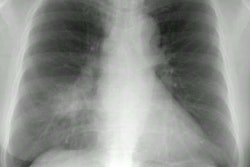
There are two predominant species that cause infection in humans- C. neoformans (infecting predominantly immunocompromised individuals) and C. gatti (which is endemic in the Northwest US and can infect both healthy and immunocompromised individuals) [7]. Birds (esp. pigeons) are reservoirs for the organism (a budding encapsulated yeast) which can be seen on stain with india ink. The organism is particularly abundant in soil contaminated by pigeon droppings. Most patients who develop cryptococcus are immune compromised or immunosuppressed- particularly HIV and Hodgkin's patients, but also patients using oral corticosteroids.Cryptococcus is the most common fungal pneumonia in HIV patients [1,3]. Most patients have severe immune compromise with CD4 counts below 100 cells/ul. Cryptococcal pneumonia is usually associated with disseminated disease and occurs in 30-45% of AIDS patients with cryptococcal infection [2,3]. The CNS is the most common site of disseminated infection- where the infection typically produces a lethal meningitis. A concomitant meningitis is found in up to 86% of cases, although it commonly clinically silent. In HIV patients that present with CNS cryptococcal infection, clinically silent pulmonary disease can be found in just over 25% of cases.
In patients with normal immunity, infection is usually
asymptomatic (one-third of patients) [5,7], although cough has
been described in up to 69% of patients [6]. Fever is uncommon (up
to 8% of patients) [6]. The most common radiographic finding is
the presence of multiple peripheral
pulmonary nodules (less than 10 mm in size [7-20mm]) which rarely
calcify. The nodules typically have well defined margins, although
irregular or poorly defined margins can also be seen [4,6]. Areas
of air space consolidation are the next most common finding and
can be seen with or without associated nodules [6]. Masses,
lymphadenopathy, and pleural effusion occur infrequently in
immunocompetent patients [4,5]. Cavitation of the nodules (or
airspace consolidation) is variable and has been reported as
infrequent [4,7] to up to 40% of cases on CT [5]. Cavitation is
more commonly seen in immunocommpromised patients [6].
Immune compromised patients present with cough (94%), fever (50%),
chest pain (44%), shortness of breath (38%), and headache (31%)
[6]. In immunocompromised patients, some authors feel that
segmental or
lobar consolidation is a common manifestation of cryptococcal
pulmonary
infection [1]. Other authors feel that interstitial infiltrates,
primarily
reticular or reticulonodular, are the most common radiographic
pattern
(50-62% of cases) [2]. Pulmonary nodules are also common and found
in up
to 30-40% of cases [1,2] and there is a propensity for the nodules
to cavitate
[1]. Nodules may be more common in patients with early HIV
infection [2].
A cavitary nodule in an HIV patient is very suggestive of either
PCP or
cryptococcal infection [1]. Hilar and mediastinal adenopathy can
also be
seen, as can pleural effusion (up to 10% of patients [6]) [1,2].
Occasionally, patients may develop
a rapidly progressive fulminant infection resulting in an ARDS
type picture
[1]. Disseminated disease typically produces a miliary pattern on
the chest
radiograph [1], although some feel that this is an atypical
presentation
for pulmonary cryptococcal infection in the HIV patient [2].
REFERENCES:
(1) Semin Roentgenology 1996; Woodring JH, et al. Pulmonary
cryptococcosis.
31 (1): 67-75 (Review. No abstract available.)
(2) Radiologic Clinics of North America 1997; McGuinness G. Changing trends in the pulmonary manifestations of AIDS. 35 (5): 1029-1082
(3) J Thorac Imaging 1999; Connolly JE, et al. Opportunistic fungal pneumonia. 14: 51-62
(4) Radiology 2005; Lindell RM, et al. Pulmonary cryptococcosis: CT findings in immunocompetent patients. 236: 326-331
(5) AJR 2005; Fox DL, Muller NL. Pulmonary cryptococcosis in immunocompetent patients: CT findings in 12 patients. 185: 622-626
(6) Chest 2006; Chang WC, et al. Pulmonary cryptococcosis.
Comparison of the clinical and radiographic characteristics in
immunocompetent and immunocompromised patients. 129: 333-340
(7) Radiographics 2021; Kunin JR, et al. Thoracic endemic fungi in the United States: importance of patient location. 41: 380-398
