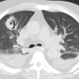
Tuberculosis (Mycobacterium tuberculosis):
View cases of TB
Clinical:
There are a number of types of TB infection:
1- Primary TB:
M. tuberculosis is an aerobic, nonmotile, non-spore forming rod
that is highly resistant to drying, acid, and alcohol [25]. The
lungs are the primary organ of spread- accounting for about 70% of
cases [27]. It is transmitted from person to person via coughing
[25]. The probability of transmission depends on the number of
infectious droplets expelled and the virulence of the strain [25].
Extrapulmonary infection generally occurs as a result of
hematogenous dissemination from a clinically occult pulmonary
focus [27]. By convention, TB is classified as primary if the
onset of clinical disease falls within one year of the initial
infection and as reactivation if the disease onset is more than
one year after initial exposure [31]. In approximately 5% of
infected individuals the immune system is inadequate at
controlling the initial infection and active TB develops (primary
infection) [32]. In another 5%, the immune system is effective at
controlling the initial infection, but viable myobacterium remain
dormant and reactivate at a later time (reactivation infection)
[32]. The remaining 90% of infected individuals never develop
symptomatic disease and will harbor infection only at a
subclinical level, which is referred to as latent TB [32]. Risk
factors associated with a higher risk of progression to active TB
include immunodeficiency (HIV is the strongest known risk factor
for developing active TB), age younger than 4 years, and IV drug
use [32].
Worldwide primary TB infection is typically seen in children (the highest prevalence in children under the age of 5 years [24]) and a large percentage of infected patients are asymptomatic [25]. In economically developed countries, the majority of cases of TB infection arise in the elderly as a result of reactivation of remote infection, or in HIV infected patients, or minorities [13]. However, due to successful treatment of the infection in the past several decades, primary infection is becoming more common in adults as this is when patients now experience their first exposure. Alcoholism is an associated factor in 30-45% of adult patients with active disease. Other risk factors include HIV, co-existent medical conditions (chronic renal failure, diabetes), and corticosteroid or other immunosuppressive therapy [13]. Idiopathic pulmonary fibrosis is also a risk factor- the incidence of TB in patients with IPF is more than 4x's higher than that in the general population [25].
Primary TB is typically a self-limited infection. About 5% of adults and up to 60% of infected children are asymptomatic [13]. Non-specific complaints of malaise, weight loss, fever, cough, or hemoptysis may be noted. Hyponatremia, caused by production of an antidiuretic hormone-like substance, may be seen in up to 11% of patients [13].
The ultimate outcome of the infection depends upon the cellular
immune response of the patient. In a patient with competent
immunity, the infection is walled off and the bacilli destroyed.
In a less competent host, the infection is walled off, but the
bacillus remains viable, but dormant for many years [25]. In
approximately 5% of infected individuals, immunity is inadequate
and clinically active disease develops within one year of
infection [13].
Primary infection typically presents as a segmental or lobar consolidation usually involving the lower lobes (although any lobe may be involved) and the appearance is often indistinguishable from bacterial pneumonia [24]. Multifocal involvement is seen in 12-24% of cases [13]. Cavitation occurs in 10-60% of cases (other authors state 29% of cases [32]). However, this classic teaching has been questioned, and other authors now suggest that the radiographic appearance of TB is independent of the time since infection and more dependent on the immune status of the infected individual [31]. Cavitary upper lobe disease (previously felt to represent reactivation) is usually seen in primary disease in immunocompetent hosts [31]. CT is more sensitive than CXR in the detection of TB infection [25].
The prevalence of lymphadenopathy is greatest in the pediatric
age group (about 83-96% of affected children [16,22,24,32]) and is
seen in about 43% of adults [24]. Unilateral adenopathy is seen in
60-80% of cases and paratracheal adenopathy in 40%. The presence
of adenopathy decreases with increasing age (it may be as low as
10% in patients in their 6th decade) [13]. Lymphadenopathy may be
the only finding on chest radiograph- particularly in infacnts
[24,32]. On CT, the enlarged nodes typically demonstrate central
low attenuation and peripheral rim enhancement with the use of
I.V. contrast [22,32], particularly if larger than 2 cm [24].
Tuberculous pleuritis occurs when a subpleural focus of Mycobacterium tuberculosis ruptures into the pleural space inciting a delayed hypersensitivity reaction [19]. It may also result from hematogeneous dissemination or by direct extension of primary disease [19]. Pleural effusion is found in 25-40% of adults, but only 5-11% of children (increasing prevalence with age) with primary infection [22,32]. Effusions can occur any time after the initial infection- but typically within 3 to 6 months [13]. The effusion is usually unilateral and on the same side as the site of initial infection [16]. Pleural fluid cultures are positive in only 20-40% of cases (pleural biopsy cultures are positive in 65-75% of cases) [13]. Pleural effusion can be the only radiographic finding indicative of primary TB infection in about 5% of cases [13,22]. Pleural effusion is less common in post primary infection (18% of cases) [32]. Tuberculous pleuritis/effusion usually resolves completely, however, it can lead to pleural thickening, calcifications, and fibrothorax [9].
Complications:
A Rasmussen (bronchial) artery aneurysm/pseudoaneurysm is a rare complication of TB infection, but it can be found in up to 5% of patients with chronic cavitary TB [20].
Bronchial stenosis occurs in 10-40% of patients with active TB [32].
Regression of radiographic findings is a slow process- requiring from 6 months to 2 years for resolution [32]. Although radiographic findings may initially worsen after institution of therapy, lack of improvement after 3 months of treatment suggests infection with a drug resistant organism [13]. The Ghon lesion represents the primary site of infection in the lungs [25]. With healing the infiltrate resolves into a small calcified pulmonary nodule or area of scarring [25]. The Ranke complex consists of the Ghon lesion plus calcified hilar nodes. Simon foci are apical nodules that are often calcified and result from hematogenous seeding at the time of initial infection [13]. Despite the presence of calcification, viable bacilli can persist [13]. Radiographic differentiation between active and inactive disease can only be made reliably on the basis of temporal evolution. The American Tuberculosis Association requires that a radiograph remain unchanged for a period of 6 months to indicate stable/inactive disease. [1,2] A marked fibrotic response is common following primary TB infection [20]. Bronchiectasis (71-86% of cases on CT) and residual cavities (12-22% of cases) are typically found in the upper lobes [13]. The hilum and mediastinum are usually retracted toward the fibrotic region [20]. Endobronchial involvement occurs in 2-4% of patients with TB and can result in bronchial stenosis [20].
A normal chest radiograph has a high negative predictive value for the presence of active TB [13]. The frequency of false negative chest radiographs is about 1% in the adult immune competent population, and between 7-15% in HIV patients [13]. Computed tomography can detect the presence of adenopathy, parenchymal consolidations, or evidence of endobronchial spread not seen on plain film radiographs.
In children, those younger than 5 years are at the highest risk for infection [23,24]. Infants tend to be more symptomatic than older children and their risk for tuberculous meningitis or miliary disease is also higher [23]. Bacterial confirmation of infection is difficult in children, and the tuberculin skin test is frequently negative in infants less than 3 months of age [23]. Common findings of infection in infants include mediastinal and hilar adenopathy (seen in 90-95% of cases [25]). The adenopathy is usually unilateral and located in the hilum or paratracheal region [25]. On CT the nodes demonstrate central necrosis with rim enhancement [25]. The adenopathy can occur with/or without airspace consolidations (consolidations are seen in about 70% of children and there is no lobar predominance [25])- especially dense, homogenenous mass-like consolidations with low attenuation areas or cavitation [23]. In children under 2 years, lobar or segmental atelectasis is frequently seen [24]. Pleural effusion is not a common feature of primary TB infection in young children and it is rare in infants [23]. Regression of radiographic findings occurs slowly [23].
2- Disseminated (Miliary) TB:
Miliary tuberculosis is a manifestation of primary infection that is seen in 1 to 7% of patients (about 3% of children and 6% of adults [22])- it is usually seen in the elderly, infants, and immunocompromised [24]. It also occurs in reactivation TB and somewhat more frequently [25]. The organism disseminates hematogenously throughout the body, not just the lungs. Miliary TB can also occur as a very rare complication of intravesical Bacille Calmette-Guerin therapy for bladder carcinoma [14]. Patients present with cough, fever, malaise, HSM, and meningitis. In the early stages of miliary disease, the CXR can be normal. Typical miliary lesions may not be visible for 3 to 6 weeks after hematogenous dissemination [16]. CXR reveals micronodular densities (1-2mm) diffusely throughout both lungs. Associated mediastinal/hilar adenopathy is common, especially in children. HRCT demonstrates a combination of sharp and poorly defined 1 to 3 mm nodules distributed throughout the lungs and have no relationship to the airways in their distribution. The nodules usually resolve within 2-6 months with treatment [24]. Healing SELDOM produces miliary calcifications.
3- Progressive Primary TB:
Widespread pulmonary infection due to impaired host immunity. Develops in approximately 5% of infected individuals [25].
4- Reactivation or Post-primary TB:
The rate of development of post-primary TB from an old primary focus is about 1% per year in persons with normal immunity (other authors suggest a lifetime risk of 10% for reactivation [31]), but it can be as high as 10% per year in patients with deficient T-cell immunity (HIV) [3,31]. Reactivation infection usually develops in the apical/posterior segments of the upper lobes (83-85% of cases) or superior segment of the lower lobes (11-14% of cases) [13]. In 70-90% of cases, the abnormalities involve more than one segment [22]. M. tuberculosis is an obligate aerobe and the predilection for the upper lung zones is likely related to the higher oxygen tension and decreased lymphatic drainage from these regions [13]. Although parenchymal infection outside of these typical locations is common, it is usually observed with associated abnormality in the characteristic sites. An atypical distribution of disease with anterior segment upper lobe or basilar segment infiltrates is found in only about 5% of cases [13].
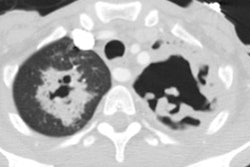
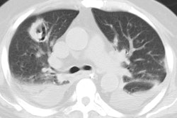
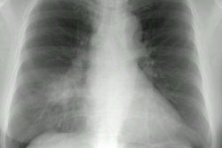
The infection usually appears as a patchy alveolar infiltrate involving the apical or posterior segments of the upper lobes, but it may cavitate in up to 50% of patients. The cavities typically have thick, irregular walls which become smooth and thin with successful treatment [24]. In about 5% of patients, reactivation manifests as a sharply marginated round, oval, or irregular lesion (also called a tuberculoma) [25]. Satellite nodules around the tuberculoma may be present in up to 80% of cases [25]. When mass-like, a tuberculoma can be mistaken for malignancy and will also be positive on FDG PET imaging [27]. Hilar or mediastinal adenopathy is unusual in reactivation TB (about 5-10% of cases [13,24,25]). Pleural effusion can be found in 15-20% of cases of post-primary TB [13,25]. Spread to other portions of the lung may occur via the bronchi - typically the lower lung zones (bronchogenic spread occurs in 20% of cases of post-primary TB). On plain film, bronchogenic spread appears as multiple ill-defined nodules distant from the site of cavity formation. Pleural effusion (about 18% of patients) is found less commonly than with primary TB and is usually small [24]. On CT, bronchogenic spread of tuberculosis commonly results in the presence of centrilobular opacities (70- 95% of cases), either nodules or branching linear structures 2-4 mm in diameter. A tree-in-bud appearance correlates with filling of the bronchioles by casseous material and peribronchial extension of the inflammatory process. This appearance can also be seen in primary infection with bronchogenic spread [4,5].
An effusion may be the sole manifestation of reactivation TB [25]. Pleural fluid adenosine deaminase (ADA) level can be used to suggest the diagnosis of TB pleurisy (elevated in TB) [25].
5- Tuberculous Airway Disease:
Tuberculous airway disease is most likely due to spread of
infection along peribronchial lymphatic channels or via direct
extension from adjacent mediastinal tuberculous lymphadenitis [7].
The infection initially results in the formation of tubercles in
the bronchial submucosa, which progress to ulceration and necrosis
of the mucosal wall. Healing occurs with variable fibrosis and
residual stenosis. TB infection is the most common cause of
inflammatory stricture of the bronchus [25]. Tracheobronchial TB
has been reported in between 2-4% to 10- 20% of patients with
primary TB [20,28]. Plain films may reveal atelectasis. CT of the
chest during active infection will reveal irregular
tracheobronchial narrowing and wall enhancement with I.V.
contrast. The mediastinal fat around the trachea often
demonstrates increased density consistent with inflammation.
During active tracheobronchial infection, associated parenchymal
lung infection is found in most cases. Associated adenopathy is
also common. Following antituberculous therapy for active disease,
airway narrowing and bronchial wall thickening will frequently
improve or normalize. Persistent, smooth narrowing of the involved
lumens (bronchial stenosis) can be seen if there has been
substantial fibrosis. [7] The left main bronchus is involved more
frequently in fibrotic disease [25]. Stenting can be used to treat
strictures associated with tuberculosis infection [28]. Stents are
typically left in place for more than 6 months (12-18 months)
[28]. Recurrent stricture following stent removal occurs in 10-30%
of patients [28]. Potential stent complications include
granulation tissue formation, mucostasis, and stent migration
[28].
6- Chronic tuberculous empyema [17]:
A chronic tuberculous empyema represents a chronic active infection of the pleural space that can be present for many years with only a paucity of clinical symptoms [19]. Patients often come to medical attention during a routine chest radiograph or after development of complications such as a bronchopleural fistula or empyema necessitatis (which occurs when the empyema extends through parietal pleura into the chest wall [20]). The pleural fluid is grossly purulent and smear positive for acid-fast bacilli. Treatment consists of pleural space drainage and anti-tuberculous chemotherapy. Unfortunately, the treatment can be complicated by the inability to re-expand trapped lung and difficulty in achieving therapeutic drug levels in the pleural fluid. The occurrence of a malignant neoplasm is a rare complication of chronic tuberculous empyema [19].
Pyothorax-associated lymphoma is a rare B-cell lymphoma that typically occurs in patients who have undergone artificial pneumothorax therapy for TB and have had a long-standing empyema (for more than 20 years) [26]. Patients typically present with chest pain, fever, and a palpable chest wall mass [26]. The incidence has been reported to be 2% in patients with chronic empyema [26].
On CXR there is usually a moderate to large loculated pleural fluid collection with pleural calcification and enlargement of the overlying ribs. CT demonstrates the loculated pleural fluid surrounded by a thick, calcified pleural rind [20].
7- Tuberculoma [18]:
A tuberculoma typically appears as a discrete nodule or mass. It results from repeated extensions of infection which result in a core of central caseous necrosis surrounded by a mantle of epitheliod cells and collagen. It can be well defined or have irregular margins and mimic a lung neoplasm. Most lesions are less than 3 cm in size and calcification can be seen in 20-30% of cases (usually ndoular or diffuse). Small satellite nodules about the larger lesion can be found in up to 80% of cases. Up to 90% of tuberculomas will demonstrate increased tracer uptake on PET FDG exams.
TB infection in HIV patients:
Clinical: The incidence of TB in the AIDS population is estimated to be 200-500 times that of the general population (other authors indicate the risk to be 50 times higher [30]) [10]. In the United States about 4-10% of HIV patients develop TB infection and it represents a reactivation of latent disease. TB infection, however, is much more prevalent in HIV patients- particularly in underdeveloped countries [8] and it is the most common opportunistic infection and leading cause of death in HIV infected patients worldwide [9]. Patients with advanced HIV disease are frequently anergic and therefore, PPD negative despite active TB infection [10]. HIV patients also have a higher incidence of negative sputum and cultures relative to the general population [10]. Treatment with a standard four-drug regimen should be instituted without delay once the diagnosis is established. Additional drugs may be required in areas with a high prevalence of multidrug resistance.
Patients with HIV and TB have a higher incidence of extrapulmonary manifestations, a higher susceptibility to latent disease reactivation, and a higher probability of developing disseminated disease than other patients [33]. Extrapulmonary TB occurs with a high incidence in HIV patients- it is found in 70% of patients with CD 4 counts below 100 cells/uL, and in 50% and 44% with CD 4 counts below 200 and 300, respectively [10]. Extrathoracic disease most frequently manifests as lymphadenitis [10].
Radiographic findings: The radiographic features of TB infection in patients with HIV depends upon their CD4 count. Early in HIV (CD4 count between 300-500 cells/uL), TB infection has a typical radiographic appearance of reactivation tuberculosis with a positive PPD, an upper lobe predominance, cavitation, effusion (20%), and adenopathy (5%), but extra-pulmonary disease can still be found in up to 20% of patients. The tuberculin skin test is positive in 40-80% of patients at this time, however, the risk of a false-negative PPD increases as the CD4 count declines. Some patients with relatively preserved immune function may not have had prior TB exposure, in those cases the infection appears as typical primary TB [10]. Between 14-20% of HIV patients with active TB may have a normal chest radiograph [6,10,12,21,29].
In patients with more advanced HIV (CD4 count between 50 and 200
cells/uL), infection often resembles primary TB with coarse
reticulonodular opacities or areas of consolidation randomly
distributed in the upper and lower lung zones. Cavitation is very
uncommon in patients with more advanced HIV infection (other
etiologies for cavitary infiltrates should also be considered in
patients with CD4 counts below 50 cells/uL including bacterial
infections with Rhodococcus equi and Nocardia
asteroides). Later, when cell mediated immunity is absent,
atypical features such as a coarse or miliary infiltrate can be
seen. Adenopathy is more common in this group of patients
(60%) and is characteristically of central low density with rim
enhancement, involving both the mediastinum and hila. TB should be
strongly suspected in any HIV patient that develops mediastinal
adenopathy [4]. Although cryptococcal infection may also produce
diffuse mediastinal adenopathy, it is usually associated with
meningeal disease. Pleural effusion is found in 4 to 13% of cases.
A miliary pattern has been reported in up to 13% of cases of TB in
HIV patients [4]. HRCT will demonstrate the presence of diffuse 1
to 3 mm nodules with associated nodular thickening of the vessels,
interlobular septa, and interlobar fissures.
Below 50 cells/uL the findings are not specific and include
diffuse consolidation, ground glass opacities, and pleural
effusion [30].
CXR findings may transiently worsen in HIV patients with active pulmonary TB between one to 5 weeks following the initiation of antiretroviral therapy [15]. This is known as immune reconstitution syndrome and reflects a delayed vigorous immune response to previous subclinical infection [32]. The most severe worsening usually occurs in patients with initially low CD4 counts (less than 50-100), but can occur even in patients with counts of more than 200 [15,32]. This phenomenon is likely related to improved immune function [15].
Extrapulmonary Tuberculosis:
Extrapulmonary TB can be seen in about 14% of cases of TB [33].
Patients with HIV and TB have a higher incidence of extrapulmonary
manifestations, a higher susceptibility to latent disease
reactivation, and a higher probability of developing disseminated
disease than other patients [33]. Extrapulmonary manifestations of
TB include: Lymphadenitis- is the most common form of
extrapulmonary TB [27]. Cervical lymphadenitis, also referred to
as scrofula, is usually caused by lymphatic spread from
mediastinal nodes after reactivation of primary lung infection
[33].
Abdominal TB is characterized by high density ascites (20-45 H.U.) and lymphadenopathy which is seen in 55-90% of cases and usually peripancreatic/mesenteric and less commonly retroperitoneal. The nodes demonstrate a low density center in 40% of cases due to necrosis. Rim calcification is NOT a feature.
Gastrointestinal involvement is most common identified in the ileocecal region (90%) of cases. Peritoneal involvement in TB is present in 5% of cases and it is usually associated with widespread abdominal disease involving lymph nodes or bowel [27]. There are three main types of TB peritoneal involvement- wet, fibrotic-fixed, and dry-plastic [27]. The fibrotic-fixed form resembles malignancy with large omental masses, matted loops of bowel, and nodular thickening of the mesentery [27].
Extrapulmonary TB frequently affects the genitourinary system,
but is usually asymptomatic [33]. Renal involvement starts in the
kidney due to hematogenous dissemination and can lead to an
autonephrectomy. The earliest imaging finding can be seen in the
minor calyces with erosion and papillary necrosis producing a
moth-eaten appearance [33]. Asymmetric caliectasis can be seen in
the more advanced stages [33]. Cortical granulomas produce solid
hypoenhancing masses in the renal parenchyma [33]. Ureteral and
bladder involvement are the result of a descending renal infection
and appear after kidney infection [33]. The ureter can appear
thickened and sometimes shows focal nodular filling defects that
represent debris or sloughed papillae [33]. Ureteral stenosis is
also frequent [33].
Skeletal TB occurs in about 3% of patients and accounts for
one-third of TB occurring in extrapulmonary locations. TB
osteomyelitis is often insidious [27]. The spine is the most
common site of skeletal involvement (about 50-60% of cases
[24,27]), followed by the hip and knee. Spinal involvement is
referred to as 'Pott's disease'. The disease typically involves
the anterior vertebral body and subligamentous spread to
contiguous vertebrae with diskitis is characteristic [27].
Involvement of three or more vertebral bodies, a kyphosis angle
greater than 25 degrees, and anterior height loss greater than 20%
are considered severe disease characteristics associated with
greater morbidity [33].
REFERENCES:
(2) ACR Syllabus #40, p.135-36
(3) RadioGraphics 1997; Rubin SA. Tuberculosis and atypical mycobacterial infections in the 1990s. 17: 1051-1059 (No abstract available)
(4) Radiol Clin North Am 1994; 32 (4): 649-661
(5) HRCT of the Lungs, 2nd ed., p.61
(6) Radiographics 1997; 17: 47-58
(8) Annu Rev Med 1996; 47: 117-26
(9) Curr Opin Pulm Med 1997; 3: 151-158
(10) Radiologic Clinics of North America 1997; McGuinness G. Changing trends in the pulmonary manifestations of AIDS. 35 (5): 1029-1082
(11) AJR 1998; Moon WK, et al. Mediastinal tuberculous lymphadenitis: CT findings of active and inactive disease. 170: 715-718
(12) J Thorac Imag 1998; Washington L, et al. Mycobacterial infection in immunocompromised patients. 13: 271-281
(13) Radiology 1999; Leung AN. Pulmonary tuberculosis: The essentials. 210: 307-322 (Review- no abstract available)
(14) AJR 1999; Rabe J, et al. Miliary tuberculosis after intravesical bacille calmette-guerin immunotherapy for carcinoma of the bladder. 172: 748-750 (No abstract available)
(15) AJR 2000; Fishman JE, et al. Pulmonary tuberculosis in AIDS patients: Transient chest radiographic worsening after initiation of antiretroviral therapy. 174: 43-49
(16) Society of Thoracic Radiology Annual Meeting 2000 Course Syllabus; Leung AN. Pulmonary tuberculosis. 83-84
(17) Semin Resp Infect 1999; Sahn SA, Iseman MD. Tuberculous empyema. 14: 82-87
(18) Radiology 2000; Goo JM, et al. Pulmonary tuberculoma evaluated by means of FDG PET: Findings in 10 cases. 216: 117-121
(19) AJR 2001; Choi JH, et al. CT manifestations of late sequelae in patients with tuberculous pleuritis. 176: 441-445 (No abstract available)
(20) Radiographics 2001; Kim HY, et al. Thoracic sequelae and complications of tuberculosis. 21: 839-860
(21) AJR 2005; Marchiori E, et al. Pulmonary disease in patients with AIDS: high-resolution CT and pathologic findings. 184: 757-764
(22) Radiol Clin N Am 2005; Tarver RD, et al. Radiology of community-acquired pneumonia. 43: 497-512
(23) AJR 2006; Kim WS, et al. Pulmonary tuberculosis in infants: radiographic and CT findings. 187: 1024-1033
(24) Radiographics 2007; Burrill J, et al. Tuberculosis: a radiologic review. 27: 1255-1273
(25) AJR 2008; Jeong YJ, Lee KS. Pulmonary tuberculosis: up-to-date imaging and management. 191: 834-844
(26) AJR 2010; Ueda T, et al. Pyothorax-associated lymphoma: imaging findings. 194: 76-84
(27) AJR 2010; Tan CH, et al. Tuberculosis: a benign impostor.
194: 555-561
(28) Radiology 2012; Verma A, et al. Posttuberculosis
tracheobronchial stenosis: use of CT to optimize the time of
silicone stent removal. 263: 562-568
(29) AJR 2012; Lichtenberger JP, et al. What a differetial a
virus makes: a practical approach to thoracic imaging findings in
the context of HIV infection- Part I, pulmonary findings. 198:
1295-1304
(30) Radiographics 2014; Chou SHS, et al. Thoracic diseases
associated with HIV infection in the era of anti-retroviral
therapy: clinical and imaging findings. 34: 895-911
(31) AJR 2015; Rozenshtein A, et al. Radiographic appearance of
pulmonary tuberculosis: dogma disapproved. 204: 974-978
(32) Radiographics 2017; Nachiappan AC, et al. Pulmonary
tuberculosis: role of radiology in diagnosis and management. 37:
52-72
(33) Radiographics 2019; Rodriguez-Takeuchi SY, et al.
Extrapulmonary tuberculosis: pathophysiology and imaging findings.
39: 2023-2037
